Lung (bottom section)
Homo sapiens
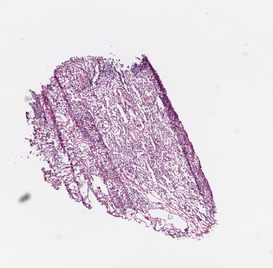
Frozen section of lung, cut from the bottom face of the frozen block. A wedge-shaped fragment of peripheral lung shows partly collapsed alveolar parenchyma: airspaces remain open through the center while the subpleural zones condense into darker basophilic bands. Fine red septal vessels trace the alveolar walls, with a more congested pocket at the lower right. Several curved section folds and marginal tears reflect the mechanics of cryosectioning aerated tissue. At higher power the septa show capillaries, flattened type I pneumocytes, and occasional plumper type II pneumocytes at septal corners.
- Category
- Histology and Tissue
- Magnification
- 20x
- Stain
- H&E
- Tags
- normal lung tissue, alveoli, alveolar septa, type I pneumocytes, type II pneumocytes, septal capillaries, subpleural atelectasis, vascular congestion, respiratory system, frozen section, tumor-adjacent normal tissue, H&E, Homo sapiens, TCGA-LUSC, TCGA-22-4596, pulmonary histology
Explore this slide with deep zoom, save it to your library, and present it live to students on StudyScope. Create a teacher account or browse the full catalog.