Lung (top section)
Homo sapiens
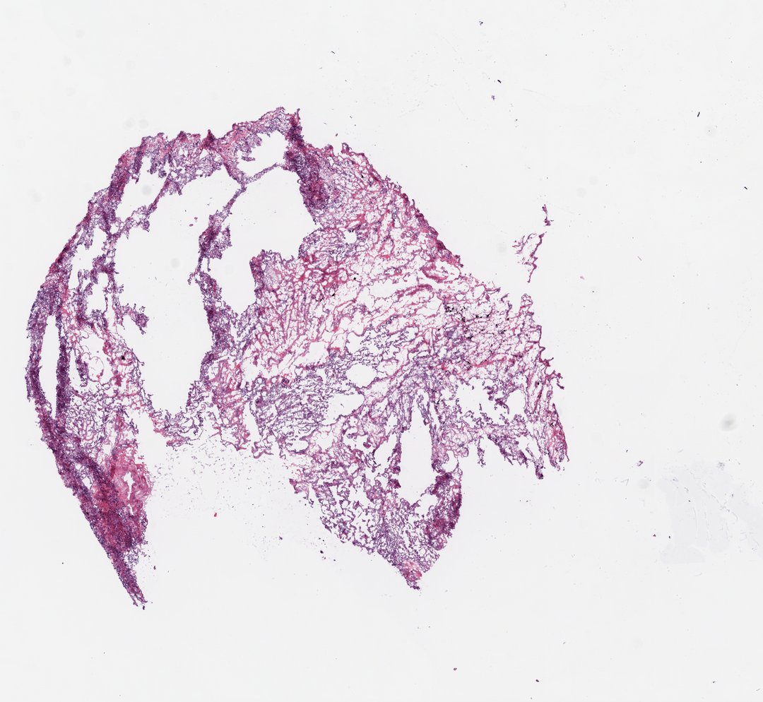
Frozen section of lung. The parenchyma forms an open, lace-like network of thin alveolar septa around widely patent airspaces — well-aerated peripheral lung at its most delicate. Denser, darker bands of collapsed alveoli follow the subpleural margin along the left of the section, and a small focus of septal congestion stains red at the lower left. The section has folded on itself in places during cryosectioning, producing the darker creases that cross the tissue; between folds the fine alveolar architecture, septal capillaries, and scattered alveolar macrophages can be followed at high power.
- Category
- Histology and Tissue
- Magnification
- 20x
- Stain
- H&E
- Tags
- normal lung tissue, alveoli, alveolar septa, pneumocytes, alveolar capillaries, subpleural atelectasis, septal congestion, alveolar macrophages, respiratory system, frozen section, section folds, tumor-adjacent normal tissue, H&E, Homo sapiens, TCGA-LUSC, TCGA-18-3414
Explore this slide with deep zoom, save it to your library, and present it live to students on StudyScope. Create a teacher account or browse the full catalog.