Onion Root Tip Mitosis Under the Microscope
Onion Root Tip Mitosis (Longitudinal Section), Allium cepa
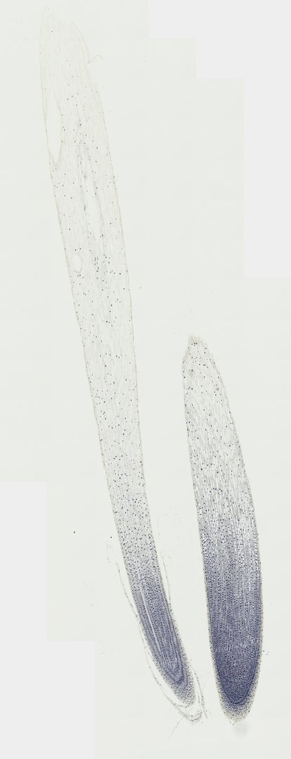
This is a lengthwise section of an onion root tip stained with iron hematoxylin. It is the classic slide for finding cells in each stage of mitosis, because root tips grow quickly and have many dividing cells.
This longitudinal section of an onion root tip (Allium cepa l.s.) captures the active regions of plant development, providing a classic model for studying somatic mitosis and root tissue zonal architecture. At the distal terminus, a protective root cap shields the apical meristem, which consists of small, densely cytoplasmic, rapidly dividing cells undergoing various stages of the cell cycle, including interphase, prophase, metaphase, anaphase, and telophase. Proximal to the meristematic zone, cells transition into the zone of elongation, where vacuolation drives longitudinal growth and distinct cell walls delineate developing protoderm, ground meristem, and procambium. Stained prominently with iron hematoxylin or acetocarmine, condensed chromatin patterns, aligned equatorial chromosomes, and separated sister chromatids stand out against the surrounding cellular walls.
What to look for
- Root cap: A cap of cells at the very tip that protects the root as it pushes through soil.
- Interphase cells: Most cells, with a round nucleus and no visible chromosomes.
- Prophase: Cells where chromosomes have thickened into visible threads inside the nucleus.
- Metaphase: Cells with chromosomes lined up across the middle.
- Anaphase: Cells with two groups of chromosomes pulling toward opposite ends.
- Telophase: Cells with two new nuclei forming and a new wall starting between them.
At each magnification
Low power (about 4x)
The rounded root tip with the root cap at the end.
Medium power (about 10x)
The zone just behind the root cap, packed with small, square cells.
High power (about 40x)
Chromosomes stained dark blue-black in cells at different stages of mitosis.
Questions and answers
What does onion root tip mitosis look like under a microscope?
In the region just behind the root cap, you will see many small, square cells. Most are in interphase with a round nucleus, but some show dark chromosomes as threads (prophase), lined up in the middle (metaphase), pulling apart (anaphase), or forming two nuclei (telophase).
Why are most onion root tip cells in interphase?
Cells spend most of their life cycle in interphase, growing and copying their DNA. Mitosis is fairly quick, so at any moment only a small share of cells are caught dividing.
Why use onion root tips to study mitosis?
Root tips grow fast, so they have many dividing cells in one small area. Onion cells also have a small number of large chromosomes (16 in a body cell), which are easy to see when stained.
- Category
- Cell Biology
- Scan magnification
- 20x
- Stain
- Iron Hematoxylin
- Tags
- onion root tip, Allium cepa, mitosis, longitudinal section, apical meristem, root cap
Viewing this slide in class
This is a scan of a real prepared slide, not a simulation or a drawing. On StudyScope you can pan from the whole slide down to individual structures, always in focus, and measure them in real micrometers with the calibrated ruler. Present it live and every student screen follows your view and annotations. Students join with a room code on Chromebooks, iPads, or any browser, with no accounts.