Short answer: To find mitosis in an onion root tip, start at the very tip and move just behind the root cap, into the zone of small, square cells. Most cells there are in interphase, with a round nucleus and no visible chromosomes. Hunt among them for the dividing cells: chromosomes as dark threads (prophase), lined up across the middle (metaphase), pulling toward opposite ends (anaphase), or forming two new nuclei with a wall starting between them (telophase). Root tips are used because they grow fast, so many cells are dividing in one small area.
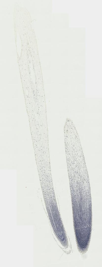 Onion Root Tip Mitosis (Longitudinal Section)Allium cepaIron Hematoxylin
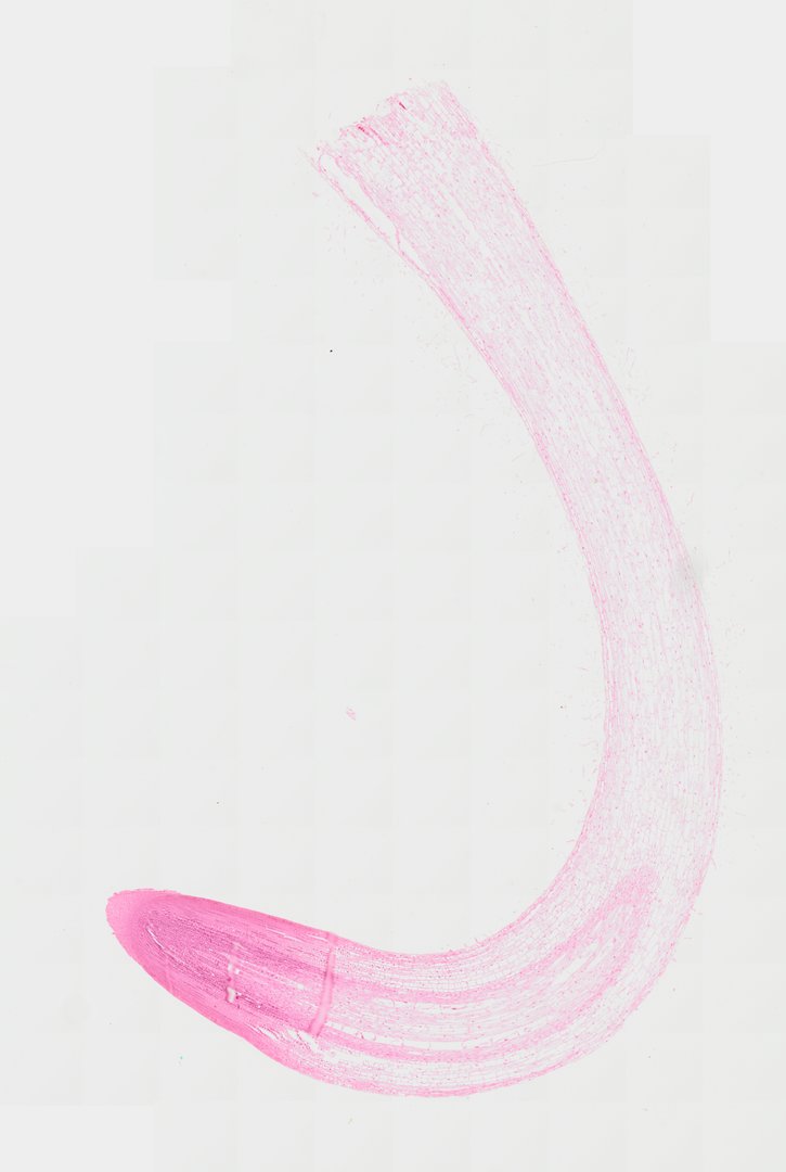 Zea Root Tip (Longitudinal Section)Zea maysSafranin
Why use the root tip to study mitosis?
A growing root makes new cells at its tip. Just behind the root cap sits the meristem, a zone of small, boxy cells that divide quickly. Farther back, cells stop dividing and stretch in the zone of elongation, which is what pushes the root deeper. Because dividing cells are crowded into one small region, a single slide can show every stage of mitosis side by side. Onion cells also have a small number of large chromosomes (16 in a body cell), which stain dark and are easy to see.
The stages of mitosis at a glance
| Stage | What you see | What is happening |
|---|---|---|
| Interphase | Round nucleus, no visible chromosomes | The cell grows and copies its DNA. This is where most cells spend most of their time. |
| Prophase | Chromosomes thickened into dark threads inside the nucleus | Chromosomes condense and become visible. |
| Metaphase | Chromosomes lined up across the middle of the cell | Chromosomes are held at the cell's center, ready to be separated. |
| Anaphase | Two groups of chromosomes pulling toward opposite ends | The copies of each chromosome are pulled apart. |
| Telophase | Two new nuclei forming, with a new wall starting between them | Nuclei re-form at each end and the cell begins to split in two. |
Labeled stages on the onion root tip
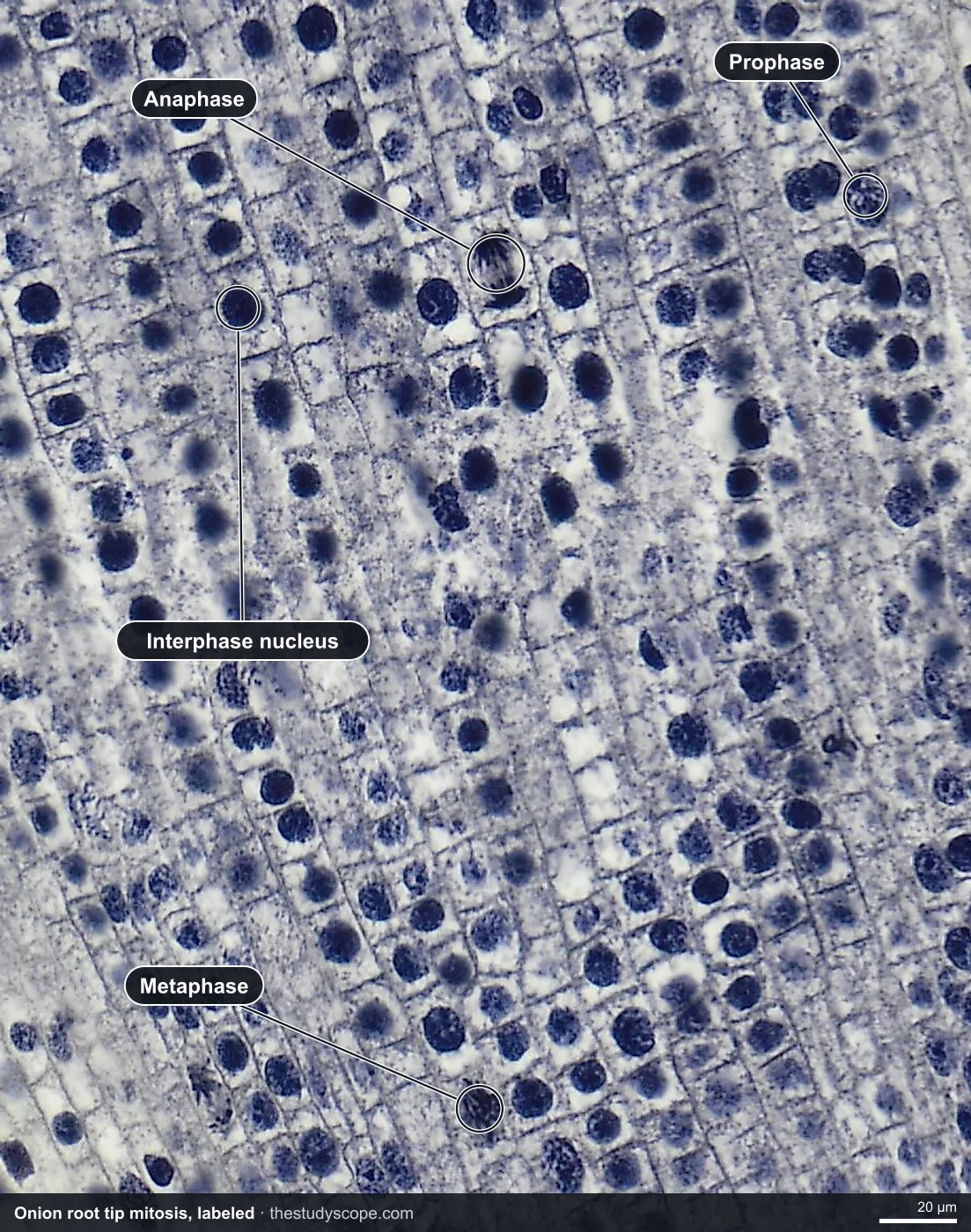
Hunting for the stages yourself is the point of the slide, but a labeled example helps you check your eye. This preview marks interphase, prophase, metaphase and anaphase. Telophase is on the slide too, so look for it on your own.
How to identify the stages of mitosis under a microscope
- Start at low power. Find the rounded tip with the root cap at the end, a cap of cells that protects the root as it pushes through soil.
- Move to medium power just behind the cap. This is the meristem, packed with small, square cells.
- Zoom in until you can see chromosomes. At high power they show as dark blue-black threads and clumps against the paler cytoplasm.
- Scan for cells that are not plain interphase. Interphase cells have a round nucleus and no visible chromosomes. Anything with visible chromosomes is dividing.
- Decide the stage by where the chromosomes are. Threads inside the nucleus means prophase, a line across the middle means metaphase, two groups moving apart means anaphase, and two new nuclei with a wall starting means telophase.
- Check a neighbor. If a cell is hard to call, find a clearer one nearby and compare. Cells caught between stages are normal.
What to look for on the onion root tip
- Root cap: the cap of cells at the very tip.
- Interphase cells: the majority, each with a round nucleus.
- Prophase: chromosomes thickened into visible threads inside the nucleus.
- Metaphase: chromosomes lined up across the middle.
- Anaphase: two groups of chromosomes pulling toward opposite ends.
- Telophase: two new nuclei forming and a new wall starting between them.
Optional: a mitotic index count
The mitotic index tells you what share of cells in an area are dividing. It turns the slide into data, and it works as a short onion root tip mitosis activity for a class.
- Pick one field of view in the meristem at high zoom, and keep it in view for the whole count.
- Count every cell in that field and sort each one into interphase, prophase, metaphase, anaphase or telophase. A tally chart works well.
- Add up all the cells you counted, and add up the cells in any stage of mitosis (everything except interphase).
- Divide the dividing cells by the total cells. Multiply by 100 for a percentage. That is the mitotic index.
- Repeat in a second field and compare. Then ask which stage had the most cells and what that suggests about how long each stage lasts.
Your numbers depend on the field you pick and how you call the in-between cells, so different students will get different counts. That is a good discussion point about sampling. Because the more cells you count, the steadier the result, pooling the whole class's fields gives a better estimate than any single count.
Onion root tip vs corn root tip
Once students can find the stages in the onion root tip, the corn (Zea mays) root tip is a good second look. It is another real longitudinal section, so the same zones appear in the same order.
| Feature | Onion root tip (Allium cepa) | Corn root tip (Zea mays) |
|---|---|---|
| Stain | Iron hematoxylin: nuclei and chromosomes dark blue | Safranin: pink to red |
| Root cap | Loose cells at the very tip | A pad of cells covering the tip |
| Meristem | Small, boxy cells, most in interphase, some in mitosis | Crowded small cells with dark nuclei, some caught in mitosis |
| Behind the meristem | Zone of elongation, cells grow longer | Zone of elongation, then tissues sorting into an outer layer, a cortex and a central core |
| Best use | Finding and counting each stage of mitosis | Seeing the zones of a root and how tissues start to form |
Onion is usually the easier slide for naming stages. Corn is a better view of the whole growing root, and it still has dividing cells with condensed chromosomes to find at high zoom.
Animal cell division in roundworm eggs
Plants and animals both divide by mitosis, but they split the cell differently. In the onion root tip, a new wall starts to form between the two new nuclei. An animal cell has no cell wall, so it pinches in two instead. The premade lesson below looks at this difference using roundworm eggs. For a first look at roundworm reproductive anatomy, open the Ascaris female cross section, where two large uterus tubes are packed with eggs beside many small ovary tubules.
Teaching it with StudyScope
- Present it live. Open the onion root tip, and students join with a room code on their own screens (no student accounts). Your annotations appear on their screens as you point out each stage.
- Let students hunt. Students can pan and zoom on their own, draw boxes around the cells they think are in each stage, and save a stamped snapshot of their view.
- Measure. Every slide is calibrated, so the ruler reads real micrometers. Students can measure a cell or the length of the meristem.
- Compare the roots. Put the onion and corn tips side by side and ask which zones match.
- Bench microscope vs virtual. A bench microscope with a wet mount lets students make their own squash and focus through a cell. A virtual slide gives every student the same well-stained section with clear stages, which is hard to guarantee at the bench.
Want it ready to teach? The premade lesson Introduction to Mitosis & Cell Division is about 35 to 40 minutes. Students hunt for the stages in an onion root tip, see how an animal cell divides in roundworm eggs, then look at a mature leaf to see why most cells are not dividing. Or open the onion root tip slide and the corn root tip slide directly.
Questions and answers
What does onion root tip mitosis look like under a microscope?
Just behind the root cap you see many small, square cells. Most are in interphase with a round nucleus. A few show dark chromosomes as threads (prophase), lined up in the middle (metaphase), pulling apart (anaphase), or forming two nuclei (telophase).
How do you identify the stages of mitosis under a microscope?
Look at where the chromosomes are. Threads inside the nucleus is prophase, a line across the middle is metaphase, two groups moving to opposite ends is anaphase, and two new nuclei with a wall starting between them is telophase. A round nucleus with no visible chromosomes is interphase.
Why are most onion root tip cells in interphase?
Cells spend most of their life cycle in interphase, growing and copying their DNA. Mitosis is fairly quick, so at any moment only a small share of cells are caught dividing.
Why is the root tip used to study mitosis?
Root tips grow fast, so the meristem just behind the root cap has many dividing cells in one small area. Onion also has a small number of large chromosomes, 16 in a body cell, which stain dark and are easy to see.
How do you calculate the mitotic index?
Count all the cells in a field of view, and count how many are in any stage of mitosis (prophase, metaphase, anaphase or telophase). Divide the dividing cells by the total cells and multiply by 100 to get a percentage. Counting more cells, or pooling several fields, gives a steadier result.
Can I do an onion root tip mitosis lab online?
Yes. StudyScope shows a real prepared onion root tip slide in the browser, on a Chromebook, iPad or computer, with deep zoom from the whole tip to single cells. Teachers can present it live and students join with a room code. A virtual slide does not replace making your own squash at the bench, but it gives every student a clear, well-stained view.
More guides
- Monocot vs dicot stems compared
- Frog, human, chicken and fish blood compared
- Virtual microscope, simulator or digital microscope?
- Using a microscope on a Chromebook
- Cheek cells vs onion cells
- Leaf layers and stomata guide
- Pond water organisms identification guide
- How to identify tissue types
- How to measure cell size
- Lily, pine and luffa pollen compared
- Insect mouthparts compared
- Worm body cavities compared