Ascaris Female Cross Section Under the Microscope
Ascaris Female (Cross Section), Ascaris lumbricoides
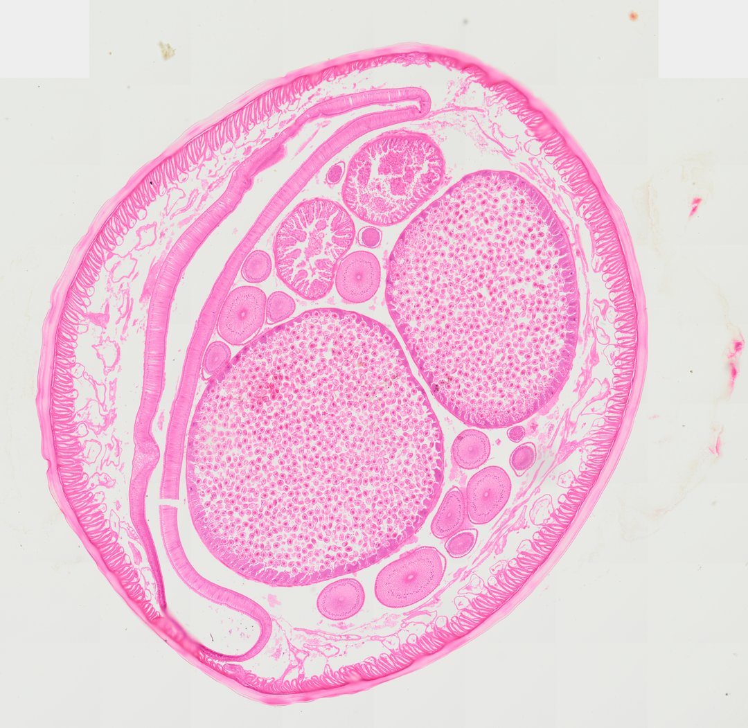
This is a cross section of a female Ascaris, a large parasitic roundworm, stained with H&E. It is a standard slide for learning roundworm body structure and comparing female and male anatomy.
A cross section of a female Ascaris roundworm displaying classic pseudocoelomate anatomy. The specimen is bounded by a prominent, non-cellular thick cuticle overlying a thin hypodermis and tall, coelomyarian muscle cells projecting into the pseudocoel. The body cavity contains paired, large, thick-walled uterine branches filled with numerous fertilized eggs/ova, cross sections of coiled ovarian tubules containing developing oocytes, and a flattened intestine lined by simple columnar epithelial cells.
What to look for
- Cuticle: A thick, smooth, pink outer layer with no cells in it, forming the worm's tough skin.
- Muscle cells: Tall muscle cells lining the inside of the body wall and bulging into the body cavity.
- Pseudocoelom: The open body cavity between the body wall and the organs.
- Uterus: Large tubes filled with many small, round eggs.
- Ovary tubules: Smaller round tubes cut many times over, packed with developing egg cells.
- Intestine: A flattened tube lined with a single layer of tall cells with purple nuclei.
At each magnification
Low power (about 4x)
A round cross section with a thick outer rim and several large and small tubes inside.
Medium power (about 10x)
Eggs packed inside the uterus, the muscle layer, and the flattened intestine.
High power (about 40x)
Individual eggs and the tall cells lining the intestine, with purple nuclei.
Questions and answers
How do you tell a female Ascaris from a male in cross section?
A female cross section has large uterus tubes stuffed with eggs, plus many small ovary tubules. A male cross section instead shows the testis and sperm duct, and no egg-filled uterus.
What is a pseudocoelom?
A pseudocoelom is a body cavity that is only partly lined by tissue from the middle germ layer. Roundworms like Ascaris have one, which is why they are called pseudocoelomates.
Why is Ascaris studied in biology class?
Ascaris is large enough to section easily and shows the basic roundworm body plan clearly. It is also a real human parasite, which connects the slide to health topics.
- Category
- Pathology & Parasitology
- Scan magnification
- 20x
- Stain
- H&E
- Tags
- Ascaris female cross section, Ascaris lumbricoides, Female roundworm, Nematoda, Pseudocoelomate, Uterus
Viewing this slide in class
This is a scan of a real prepared slide, not a simulation or a drawing. On StudyScope you can pan from the whole slide down to individual structures, always in focus, and measure them in real micrometers with the calibrated ruler. Present it live and every student screen follows your view and annotations. Students join with a room code on Chromebooks, iPads, or any browser, with no accounts.