Taenia Pisiformis Cross Section Under the Microscope
Taenia Pisiformis (Cross Section), Taenia pisiformis
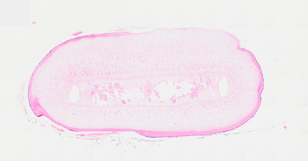
This is a cross section through a segment (proglottid) of the tapeworm Taenia pisiformis, stained with H&E. Tapeworms have no mouth or gut, so this slide shows how a parasite can be built around reproduction.
A cross section of a mature proglottid from the tapeworm Taenia pisiformis. The outer tegument displays a specialized microtriche-lined surface boundary surrounding a fibrous subtegumentary layer. The central parenchymal core contains muscular fibers, paired excretory canals, and internal reproductive tract structures including follicular testes and vitelline elements stained in shades of pink and eosin, characteristic of flatworm (Cestoda) morphology.
What to look for
- Tegument: The outer covering of the worm, through which it absorbs food.
- Muscle layers: Bands of muscle just under the tegument, stained pink.
- Parenchyma: Loose tissue filling the inside of the segment.
- Excretory canals: Small round canals running along the sides of the worm.
- Reproductive organs: Structures such as testes and yolk glands filling much of the segment.
At each magnification
Low power (about 4x)
A flat oval section with a tissue-filled center and no gut.
Medium power (about 10x)
The outer layers, the excretory canals, and the reproductive organs inside.
High power (about 40x)
Cells of the reproductive organs and the texture of the tegument.
Questions and answers
Do tapeworms have a digestive system?
No. Tapeworms have no mouth or gut. They live in their host's intestine and absorb already-digested food directly through their outer covering, the tegument.
What is a proglottid?
A proglottid is one segment of a tapeworm's body. Each mature segment contains both male and female reproductive organs, and ripe segments full of eggs break off and leave the host.
What animals does Taenia pisiformis infect?
Taenia pisiformis is a tapeworm of dogs and related animals. Rabbits and hares are its intermediate hosts, and dogs get infected by eating them.
- Category
- Pathology & Parasitology
- Scan magnification
- 20x
- Stain
- H&E
- Tags
- Taenia pisiformis cross section, Taenia pisiformis, Cestoda, Tapeworm, Proglottid, Tegument
Viewing this slide in class
This is a scan of a real prepared slide, not a simulation or a drawing. On StudyScope you can pan from the whole slide down to individual structures, always in focus, and measure them in real micrometers with the calibrated ruler. Present it live and every student screen follows your view and annotations. Students join with a room code on Chromebooks, iPads, or any browser, with no accounts.