Ascaris Male Cross Section Under the Microscope
Ascaris Male (Cross Section), Ascaris lumbricoides
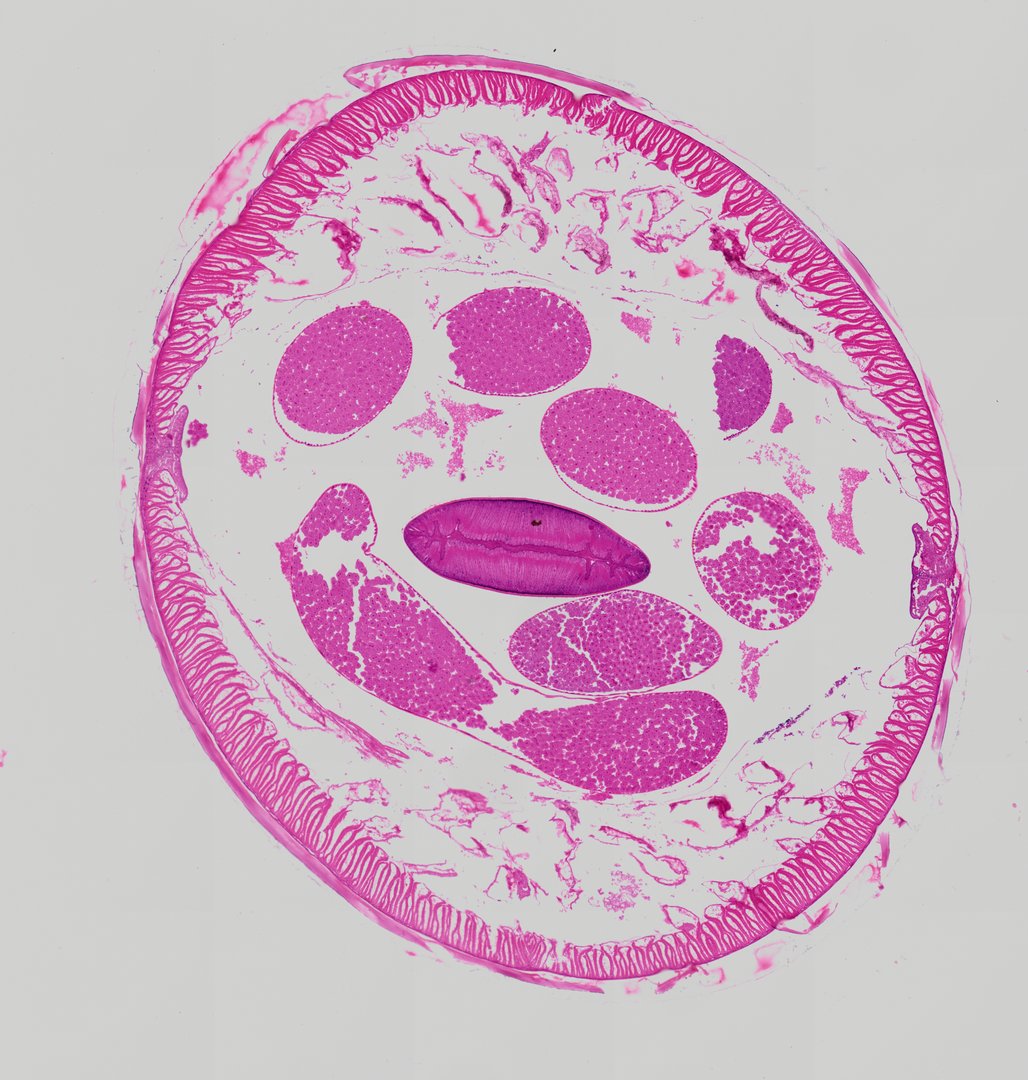
This is a cross section of a male Ascaris roundworm, stained with H&E. It shows the roundworm body plan and the male reproductive tubes, and is often compared side by side with the female slide.
A cross section of a male Ascaris roundworm displaying pseudocoelomate internal organization. The outer body wall consists of a thick, non-cellular cuticle overlying a thin hypodermis and prominent polymyarian coelomyarian longitudinal muscle cells protruding into the body cavity. Within the pseudocoelom, multiple sections of the tubular male reproductive tract—including sections of the testis filled with developing spermatocytes and the vas deferens—are visible surrounding a centrally located, flattened intestine with columnar epithelial lining.
What to look for
- Cuticle: A thick, pink, non-cellular outer layer around the whole section.
- Muscle cells: Muscle cells lining the body wall and projecting into the body cavity.
- Pseudocoelom: The open body cavity surrounding the internal tubes.
- Testis: Small round tubes, cut several times, filled with developing sperm cells.
- Vas deferens: A larger tube that carries sperm toward the rear of the worm.
- Intestine: A flattened tube lined by a single layer of tall cells.
At each magnification
Low power (about 4x)
A round section with a thick rim and a cluster of tubes inside.
Medium power (about 10x)
The muscle layer, the flattened intestine, and the sections of reproductive tubes.
High power (about 40x)
Tightly packed developing sperm cells inside the testis tubes, with purple-stained nuclei.
Questions and answers
What does a male Ascaris cross section look like under a microscope?
It shows a thick outer cuticle, a layer of muscle cells, a body cavity, a flattened intestine, and several small tubes of the male reproductive system. Unlike the female, there is no uterus full of eggs.
Are male Ascaris smaller than females?
Yes. Male Ascaris are usually noticeably smaller than females, and the male tail is curled. The curl cannot be seen in a cross section, but the smaller diameter and reproductive tubes help tell them apart.
- Category
- Pathology & Parasitology
- Scan magnification
- 20x
- Stain
- H&E
- Tags
- Ascaris male cross section, Ascaris lumbricoides, Male roundworm, Nematoda, Pseudocoelomate, Cuticle
Viewing this slide in class
This is a scan of a real prepared slide, not a simulation or a drawing. On StudyScope you can pan from the whole slide down to individual structures, always in focus, and measure them in real micrometers with the calibrated ruler. Present it live and every student screen follows your view and annotations. Students join with a room code on Chromebooks, iPads, or any browser, with no accounts.