Mushroom Gills Cross Section Under the Microscope
Mushroom Gills (Cross Section), Agaricus
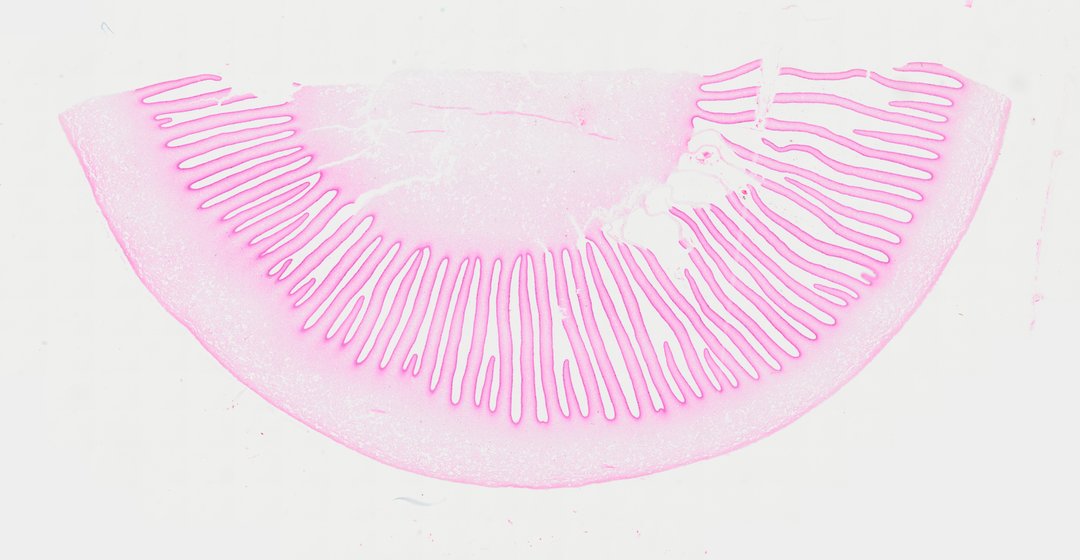
This is a section through the cap and gills of a mushroom, stained with safranin. It shows where mushrooms make their spores, on tiny club-shaped cells lining the gills.
A cross section through the cap and gills of a mushroom fruiting body (Mushroom sec) stained pink/red. The central cap tissue (pileus) supports numerous radiating gills (lamellae) hanging vertically beneath it. Microscopic examination of the gill surfaces reveals the fertile hymenium layer, where club-shaped basidia produce unicellular haploid basidiospores mounted on delicate sterigmata. The interior of each gill consists of interwoven fungal hyphae forming the central trama. This slide provides a classic demonstration of basidiomycete reproductive structures and spore-bearing hymenial anatomy.
What to look for
- Cap (pileus): The tissue at the top that holds the gills.
- Gills (lamellae): Thin plates hanging down from the cap in rows.
- Hymenium: The layer of fertile cells along the surface of each gill.
- Basidia: Club-shaped cells in the hymenium that produce spores.
- Basidiospores: Tiny spores sitting on small points at the tips of the basidia.
- Trama: Woven threads (hyphae) in the middle of each gill.
At each magnification
Low power (about 4x)
Rows of gills hanging from the cap, stained pink to red.
Medium power (about 10x)
The dense outer layer of each gill and the looser core.
High power (about 40x)
Club-shaped basidia with small spores at their tips.
Questions and answers
What do mushroom gills look like under a microscope?
Mushroom gills are thin plates with a core of woven fungal threads. Their surfaces are lined with club-shaped cells called basidia, each carrying a few tiny spores at its tip.
How do mushrooms reproduce?
Mushrooms release huge numbers of spores from their gills. The spores are carried by air, and if one lands in a good spot, it grows into new fungal threads that can eventually form a new mushroom.
Are mushrooms plants?
No. Mushrooms are fungi. They cannot make their own food by photosynthesis and instead absorb nutrients from their surroundings, and their cell walls contain chitin instead of cellulose.
- Category
- Microbiology
- Scan magnification
- 20x
- Stain
- Safranin
- Tags
- mushroom cross section, Mushroom sec, Agaricus, gills, lamellae, pileus
Viewing this slide in class
This is a scan of a real prepared slide, not a simulation or a drawing. On StudyScope you can pan from the whole slide down to individual structures, always in focus, and measure them in real micrometers with the calibrated ruler. Present it live and every student screen follows your view and annotations. Students join with a room code on Chromebooks, iPads, or any browser, with no accounts.