Euglena Under the Microscope
Euglena (Whole Mount), Euglena
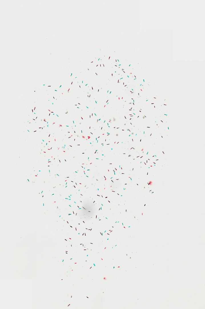
This slide shows many Euglena cells, single-celled organisms that can both photosynthesize and take in food. Euglena is a classic example of a protist that does not fit neatly into plant or animal groups.
A whole mount preparation of Euglena displaying individual unicellular flagellates with characteristic elongated, spindle-shaped morphology. The stained sample highlights single-celled protists dispersed across the field, showcasing cellular features such as the pellicle and dense cytoplasmic contents typical of mixotrophic euglenids.
What to look for
- Spindle-shaped cells: Long cells that are wide in the middle and taper toward the ends.
- Pellicle: A flexible outer covering that lets the cell bend and change shape.
- Chloroplasts: Small bodies inside the cell used for photosynthesis.
- Nucleus: A round structure near the middle of the cell.
- Flagellum: A long whip-like thread at the front end, often hard to see on prepared slides.
At each magnification
Low power (about 4x)
Small scattered specks across the slide.
Medium power (about 10x)
Individual cells with their long, tapered shape.
High power (about 40x)
Internal contents of the cell, such as the nucleus and chloroplasts.
Questions and answers
What does Euglena look like under a microscope?
Euglena is a single cell shaped like a spindle, often around 50 micrometers long. In living cells you can see green chloroplasts, a red eyespot near the front, and a long flagellum it uses to swim.
Is Euglena a plant or an animal?
Euglena is a protist. It can make food by photosynthesis like a plant, but it can also take in food from its surroundings and swims with a flagellum, which are animal-like traits.
- Category
- Microbiology
- Scan magnification
- 20x
- Tags
- Euglena, Euglena whole mount, Euglenida, Euglenaceae, Unicellular organism, Flagellate
Viewing this slide in class
This is a scan of a real prepared slide, not a simulation or a drawing. On StudyScope you can pan from the whole slide down to individual structures, always in focus, and measure them in real micrometers with the calibrated ruler. Present it live and every student screen follows your view and annotations. Students join with a room code on Chromebooks, iPads, or any browser, with no accounts.