Rooster Comb Cross Section Under the Microscope
Rooster Comb (Cross Section), Gallus gallus domesticus
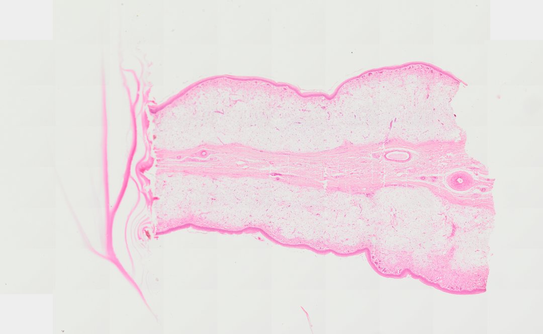
This is a cross section of a rooster comb stained with H&E. The comb is a fleshy part of the skin with many blood vessels, which is what makes it red.
A cross section of a rooster comb (c.s.) stained with hematoxylin and eosin (H&E), illustrating the complex integumentary structure unique to avian fleshy protuberances. The outermost layer is lined by a keratinized stratified squamous epithelium supported by a dense dermal layer. Beneath this lies a highly specialized, mucoid central dermis (core) rich in extracellular matrix components, notably hyaluronic acid, interspersed with a dense network of prominent capillaries, arterioles, and venules responsible for vascular turgescence, thermoregulation, and secondary sexual display.
What to look for
- Epidermis: A thin outer layer of stacked cells covering the comb.
- Dermis: Pink connective tissue under the epidermis.
- Blood vessels: Many small vessels and capillaries in the dermis, some filled with red blood cells.
- Central core: A pale, loose, jelly-like layer in the middle of the comb.
At each magnification
Low power (about 4x)
A section with a thin outer layer and a pale core, with many small vessels.
Medium power (about 10x)
The dense network of blood vessels under the skin.
High power (about 40x)
Red blood cells inside the capillaries and the loose tissue of the core.
Questions and answers
Why is a rooster comb red?
A rooster comb has a dense network of blood vessels close to the surface, and the blood shows through the thin skin. The color is part of how roosters signal health to other chickens.
What is a rooster comb made of?
A rooster comb is skin with a thin outer layer, a dermis packed with blood vessels, and a jelly-like core. That core is rich in a substance called hyaluronic acid.
- Category
- Histology and Tissue
- Scan magnification
- 20x
- Stain
- H&E
- Tags
- rooster comb, Gallus gallus domesticus, comb cross section, avian histology, stratified squamous epithelium, mucoid connective tissue
Viewing this slide in class
This is a scan of a real prepared slide, not a simulation or a drawing. On StudyScope you can pan from the whole slide down to individual structures, always in focus, and measure them in real micrometers with the calibrated ruler. Present it live and every student screen follows your view and annotations. Students join with a room code on Chromebooks, iPads, or any browser, with no accounts.
More Histology and Tissue slides
- Bone Cell (Hyaline Cartilage / Trachea Section)
- Lung (bottom section)
- Lung (bottom section)
- Lung (top section)
- Lung (top section)
- Lung (top section)
- Lung (top section)
- Oral Epithelial Cells (Whole Mount)