Bone Cell Under the Microscope
Bone Cell (Hyaline Cartilage / Trachea Section), Homo sapiens
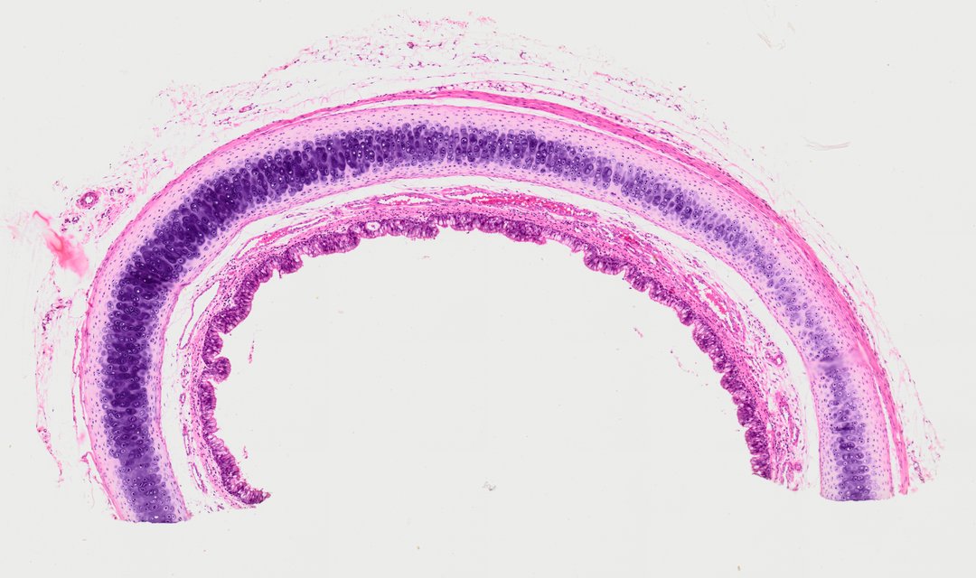
Although it is labeled as a bone cell slide, this section is really a cross section through the trachea (windpipe) showing hyaline cartilage, stained with H&E. It is useful for seeing cartilage cells and the lining of the airway.
A cross section of a tracheal C-shaped ring featuring hyaline cartilage tissue (labeled broadly as bone cell sec.). The section displays the inner pseudostratified ciliated columnar epithelium, submucosa, and the prominent avascular hyaline cartilage layer containing chondrocytes housed within lacunae embedded in a basophilic extracellular matrix.
What to look for
- Hyaline cartilage: A wide, smooth band of matrix, stained light purple to bluish, that forms the C-shaped tracheal ring.
- Chondrocytes: Cartilage cells sitting in small spaces scattered through the matrix, sometimes in pairs or small groups.
- Lacunae: The small pockets that hold the chondrocytes, sometimes showing a clear ring around the cell.
- Pseudostratified ciliated columnar epithelium: The inner lining of the airway, with nuclei at different heights and a fringe of cilia on top.
- Submucosa: Pink connective tissue between the lining and the cartilage, which may include small glands.
At each magnification
Low power (about 4x)
The curved cartilage band and the airway lining along one edge.
Medium power (about 10x)
Chondrocytes scattered in the cartilage and the layers between cartilage and lining.
High power (about 40x)
Individual chondrocytes in their lacunae and the cilia on the lining cells.
Questions and answers
What does hyaline cartilage look like under a microscope?
Hyaline cartilage looks like a smooth, glassy background with small cells (chondrocytes) sitting in little pockets called lacunae. It has no blood vessels, and the cells often sit in pairs or small groups.
Is this slide bone or cartilage?
It is cartilage. Bone has hard, layered rings (osteons) with cells in tiny spaces connected by channels, while this slide shows the smooth matrix and scattered cells of hyaline cartilage from the trachea.
Why does the trachea have cartilage?
The C-shaped rings of hyaline cartilage keep the trachea open so air can flow in and out. The open part of the C lets the esophagus behind it expand when you swallow.
- Category
- Histology and Tissue
- Scan magnification
- 20x
- Stain
- Hematoxylin and eosin
- Tags
- Bone cell sec., Hyaline cartilage section, Trachea section, Hyaline cartilage, Chondrocytes, Lacunae
Viewing this slide in class
This is a scan of a real prepared slide, not a simulation or a drawing. On StudyScope you can pan from the whole slide down to individual structures, always in focus, and measure them in real micrometers with the calibrated ruler. Present it live and every student screen follows your view and annotations. Students join with a room code on Chromebooks, iPads, or any browser, with no accounts.
More Histology and Tissue slides
- Lung (bottom section)
- Lung (bottom section)
- Lung (top section)
- Lung (top section)
- Lung (top section)
- Lung (top section)
- Oral Epithelial Cells (Whole Mount)
- Ovary (top section)