Oral Epithelial Cells Under the Microscope
Oral Epithelial Cells (Whole Mount), Homo sapiens
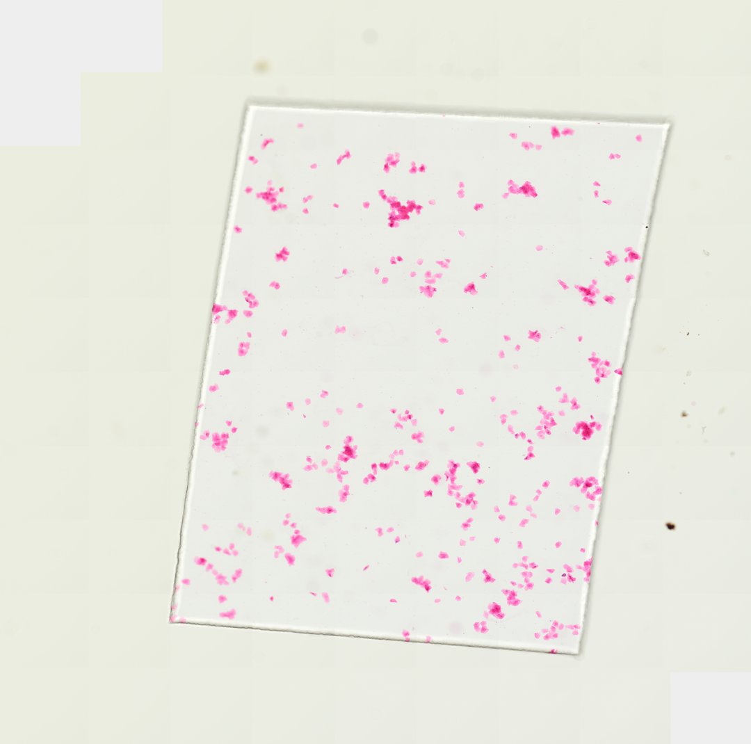
This is a smear of human cheek cells stained pink with eosin. It is a classic first look at animal cells, often compared with onion cells to show the difference between plant and animal cells.
A whole mount (W.M.) smear of human oral epithelial cells (buccal mucosa), stained pink for clarity. The preparation shows scattered flat, polygonal stratified squamous cells with prominent central nuclei and abundant cytoplasm, typical of human mucosal epithelium.
What to look for
- Cheek cells: Flat, irregular cells with many sides, spread out on the slide.
- Nucleus: A small round to oval body near the center of each cell, appearing darker than the cytoplasm.
- Cytoplasm: The pink-stained material filling most of each cell.
- Cell membrane: The thin outer edge of each cell, with no cell wall.
- Folds and clumps: Some cells may be folded or overlapping, since they are thin and flexible.
At each magnification
Low power (about 4x)
Scattered pink flakes across the slide.
Medium power (about 10x)
Individual flat cells, each with a small nucleus.
High power (about 40x)
The outline of the cell membrane and the nucleus inside.
Questions and answers
What do cheek cells look like under a microscope?
Cheek cells are flat, irregular cells with a small round nucleus near the middle. They have no cell wall, so their edges are thin and they can fold or wrinkle.
What is the difference between cheek cells and onion cells?
Cheek cells are animal cells: they have no cell wall, no large central vacuole, and an irregular shape. Onion cells are plant cells, with a rigid cell wall, a boxy shape, and a big vacuole.
- Category
- Histology and Tissue
- Scan magnification
- 20x
- Stain
- Eosin
- Tags
- Oral epithelial cells whole mount, Oral epithelial cells W.M., Buccal mucosa, Buccal epithelial cells, Stratified squamous epithelium, Human epithelial cells
Viewing this slide in class
This is a scan of a real prepared slide, not a simulation or a drawing. On StudyScope you can pan from the whole slide down to individual structures, always in focus, and measure them in real micrometers with the calibrated ruler. Present it live and every student screen follows your view and annotations. Students join with a room code on Chromebooks, iPads, or any browser, with no accounts.
More Histology and Tissue slides
- Bone Cell (Hyaline Cartilage / Trachea Section)
- Lung (bottom section)
- Lung (bottom section)
- Lung (top section)
- Lung (top section)
- Lung (top section)
- Lung (top section)
- Ovary (top section)