Lung Under the Microscope (top section)
Lung (top section), Homo sapiens
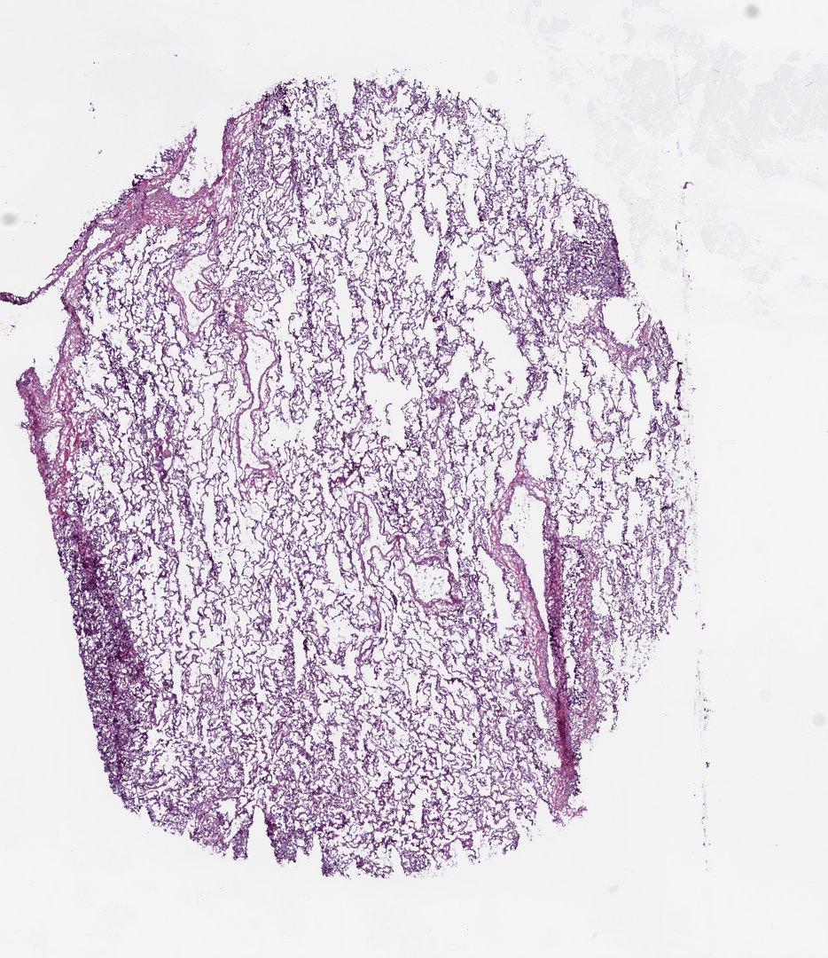
This is a frozen section of human lung tissue stained with H&E. It shows the tiny air sacs (alveoli) where oxygen passes into the blood, and it also shows the tears and folds that often happen when soft, airy tissue is frozen and cut.
Frozen section of lung. A rounded, well-filled section of peripheral lung shows remarkably even alveolar architecture: a fine honeycomb of thin septa and open airspaces occupying nearly the whole fragment. Along the left border the parenchyma darkens into a rim of peripheral atelectasis with septal congestion, and gentle vertical folds from the cryostat blade cross the tissue face. The uniformity of this section makes it a good baseline reference for normal aerated alveolar lung against which the more congested, collapsed, or emphysematous sections in this series can be compared.
What to look for
- Alveoli: Many small, open, empty-looking spaces that give lung tissue a lacy, honeycomb look.
- Alveolar walls (septa): Very thin pink walls between the air spaces, dotted with purple nuclei.
- Blood vessels: Tubes of various sizes, some filled with bright pink to red blood cells.
- Collapsed areas: Darker, denser patches where the air spaces have closed up, often near the edges.
- Folds and tears: Darker creases and gaps caused by cutting frozen tissue, not by the lung itself.
At each magnification
Low power (about 4x)
A lacy pink network with lots of open white space, plus darker bands and folds.
Medium power (about 10x)
The pattern of alveoli and the blood vessels running between them.
High power (about 40x)
Thin alveolar walls with flattened cells, purple nuclei, and red blood cells in tiny capillaries.
Questions and answers
What does lung tissue look like under a microscope?
Lung tissue looks like lace or a sponge. Most of it is tiny air sacs called alveoli, separated by very thin walls. With H&E stain, the walls are pink with purple nuclei, and blood in the vessels is red.
Why are alveolar walls so thin?
Oxygen and carbon dioxide move between the air and the blood by diffusion, which only works well over very short distances. The thin walls put the blood in tiny capillaries very close to the air.
What is a frozen section?
A frozen section is made by freezing tissue and slicing it right away instead of embedding it in wax. It is fast, but soft or airy tissue like lung often tears or folds, so some of the gaps and creases on this slide come from the cutting process.
Based upon data generated by the TCGA Research Network (https://www.cancer.gov/tcga) and made available through the NCI Genomic Data Commons (GDC). See credits.
- Category
- Histology and Tissue
- Scan magnification
- 20x
- Stain
- H&E
- Tags
- normal lung tissue, alveoli, alveolar septa, pneumocytes, alveolar architecture, peripheral atelectasis
Viewing this slide in class
This is a scan of a real prepared slide, not a simulation or a drawing. On StudyScope you can pan from the whole slide down to individual structures, always in focus, and measure them in real micrometers with the calibrated ruler. Present it live and every student screen follows your view and annotations. Students join with a room code on Chromebooks, iPads, or any browser, with no accounts.
More Histology and Tissue slides
- Bone Cell (Hyaline Cartilage / Trachea Section)
- Lung (bottom section)
- Lung (bottom section)
- Lung (top section)
- Lung (top section)
- Lung (top section)
- Oral Epithelial Cells (Whole Mount)
- Ovary (top section)