Ovary Under the Microscope (top section)
Ovary (top section), Homo sapiens
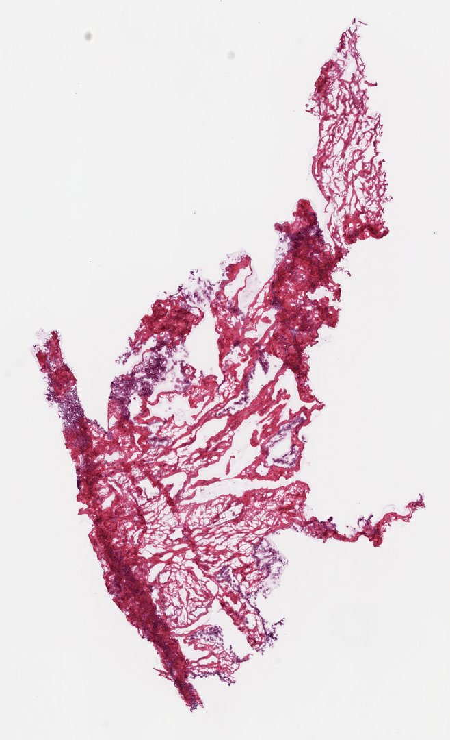
This is a frozen section of human ovary tissue stained with H&E. It mostly shows the ovary's supporting tissue (stroma) and blood vessels.
Frozen section of ovary. The section is dominated by densely collagenized ovarian stroma: whorled fascicles of spindle-shaped stromal cells within an eosinophilic collagen matrix, the fibrotic cortical pattern typical of the postmenopausal ovary. Prominent congested medullary blood vessels track through the deeper stroma, and scattered basophilic cellular foci punctuate the fibrous background. No developing follicles are conspicuous at scanning power. Cryosectioning has produced characteristic tissue tearing and fragmentation along the stromal planes, a useful example of frozen-section artifact in a collagen-rich organ.
What to look for
- Stroma: Dense tissue of spindle-shaped cells with purple nuclei, set in pink collagen, often in swirls.
- Blood vessels: Tubes of various sizes running through the stroma, some filled with red blood cells.
- Collagen: Pink fibers between the stromal cells.
- Tears: Gaps and cracks in the tissue caused by cutting frozen tissue.
At each magnification
Low power (about 4x)
A section of dense pink and purple tissue with some gaps and tears.
Medium power (about 10x)
Swirls of stromal cells and the blood vessels between them.
High power (about 40x)
The long, thin nuclei of stromal cells and blood cells inside vessels.
Questions and answers
What does ovary tissue look like under a microscope?
Ovary tissue has a dense background of spindle-shaped cells and collagen called stroma, with blood vessels running through it. In younger ovaries, you may also see round follicles, each holding an egg cell.
Why are there no follicles on some ovary slides?
The number of follicles drops with age, and after menopause the ovary is mostly stroma. Any single section is also a very thin slice, so it may miss the follicles that are present.
Based upon data generated by the TCGA Research Network (https://www.cancer.gov/tcga) and made available through the NCI Genomic Data Commons (GDC). See credits.
- Category
- Histology and Tissue
- Scan magnification
- 20x
- Stain
- H&E
- Tags
- normal ovary, ovarian stroma, ovarian cortex, stromal spindle cells, collagen, medullary vessels
Viewing this slide in class
This is a scan of a real prepared slide, not a simulation or a drawing. On StudyScope you can pan from the whole slide down to individual structures, always in focus, and measure them in real micrometers with the calibrated ruler. Present it live and every student screen follows your view and annotations. Students join with a room code on Chromebooks, iPads, or any browser, with no accounts.
More Histology and Tissue slides
- Bone Cell (Hyaline Cartilage / Trachea Section)
- Lung (bottom section)
- Lung (bottom section)
- Lung (top section)
- Lung (top section)
- Lung (top section)
- Lung (top section)
- Oral Epithelial Cells (Whole Mount)