Short answer: To read a histology slide, start at the lowest zoom and look at the layout before any single cell: find the edges, any open spaces (lumens), and the layers. Then use the stain, since in H&E nuclei are purple and most other tissue is pink. Last, zoom in to the cells. Epithelium is tightly packed cells forming a lining or covering. Connective tissue is cells scattered in a lot of material between them, like the glassy matrix of cartilage. Muscle is long fibers, and skeletal muscle shows fine stripes and nuclei at the edge of each fiber.
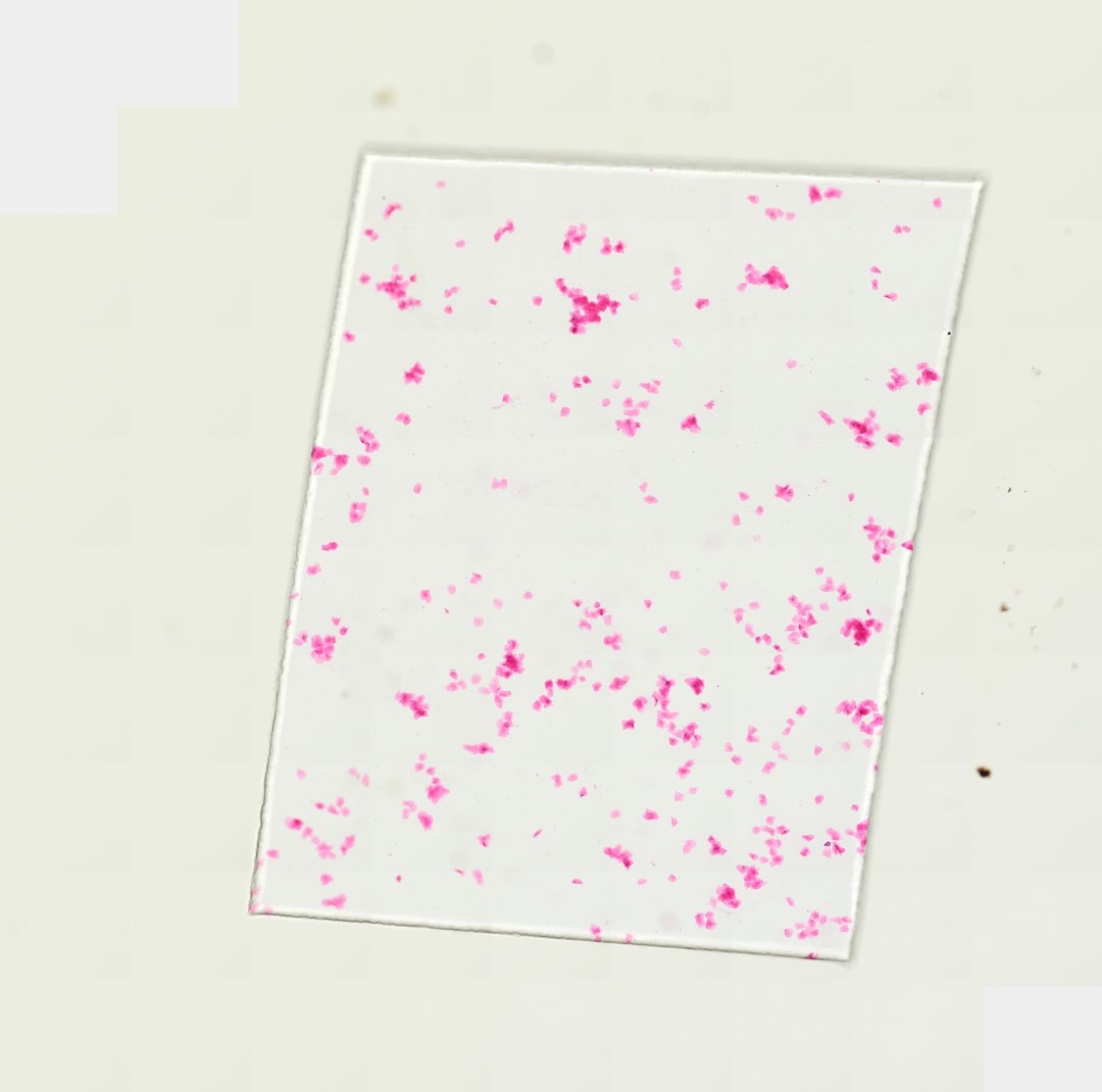 Human Cheek Cells (Whole Mount)Homo sapiensEosin
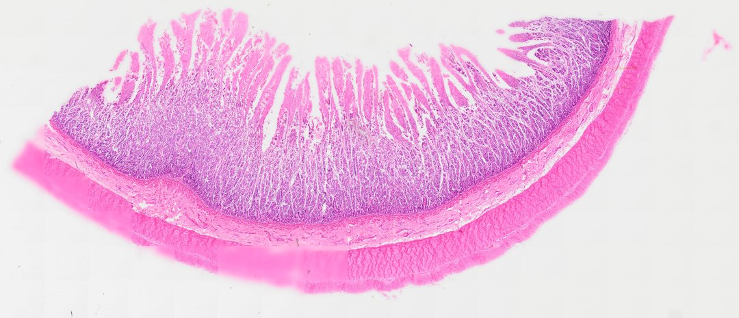 Small Intestine (Cross Section)H&E
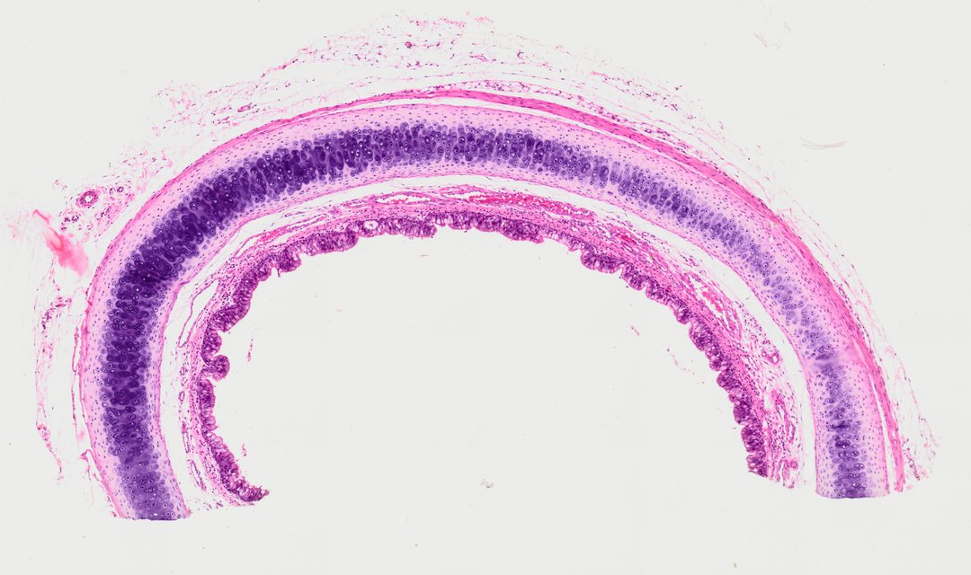 Hyaline Cartilage (Trachea Section)Homo sapiensHematoxylin and eosin
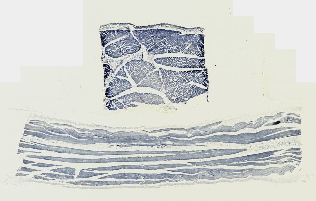 Skeletal Muscle (Transverse & Longitudinal Section)Homo sapiensIron hematoxylin
How to read a histology slide, step by step
- Start at the lowest zoom. Look at the whole section first. What is the overall shape? Is it a ring, a strip, a lacy network, or a solid piece?
- Find the edges, open spaces, and layers. A free surface with cells lined up along it points to epithelium. An open space in the middle is a lumen, as in the small intestine. Layers stacked from the lumen outward, like lining, then connective tissue, then muscle, tell you which organ you are in.
- Check the stain. In H&E (hematoxylin and eosin), nuclei stain purple and most other tissue stains pink. A smooth, glassy purple to bluish band, like the cartilage in the trachea, is matrix rather than cells. Not every slide uses H&E: the cheek cells here are stained with eosin, the skeletal muscle with iron hematoxylin, and the blood smear with Wright-Giemsa. Read the stain on the slide page before you decide what a color means.
- Zoom in to cells and nuclei. Are the cells packed edge to edge, or scattered in empty-looking material? Are they flat, tall, round, or long? Where are the nuclei: in the middle, at the edge, or at different heights?
- Name the tissue, then the organ. Decide epithelial, connective, or muscle first. Once you know the tissues, the layers tell you the organ.
Tissue types under the microscope, and a slide for each
| Tissue type | What it looks like | Slide to open |
|---|---|---|
| Epithelial | Cells packed together as a lining or covering. Cheek cells are flat with a small nucleus near the middle. The small intestine lining is a single layer of tall cells with pale mucus-making goblet cells. | [Oral epithelial cells](/catalog/slide/oral-epithelial-cells), [small intestine](/catalog/slide/small-intestine-cross-section) |
| Connective: cartilage | Small cells (chondrocytes) in little pockets (lacunae) set in a smooth, glassy matrix, with no blood vessels. | [Hyaline cartilage, trachea](/catalog/slide/bone-cell-hyaline-cartilage-trachea-section) |
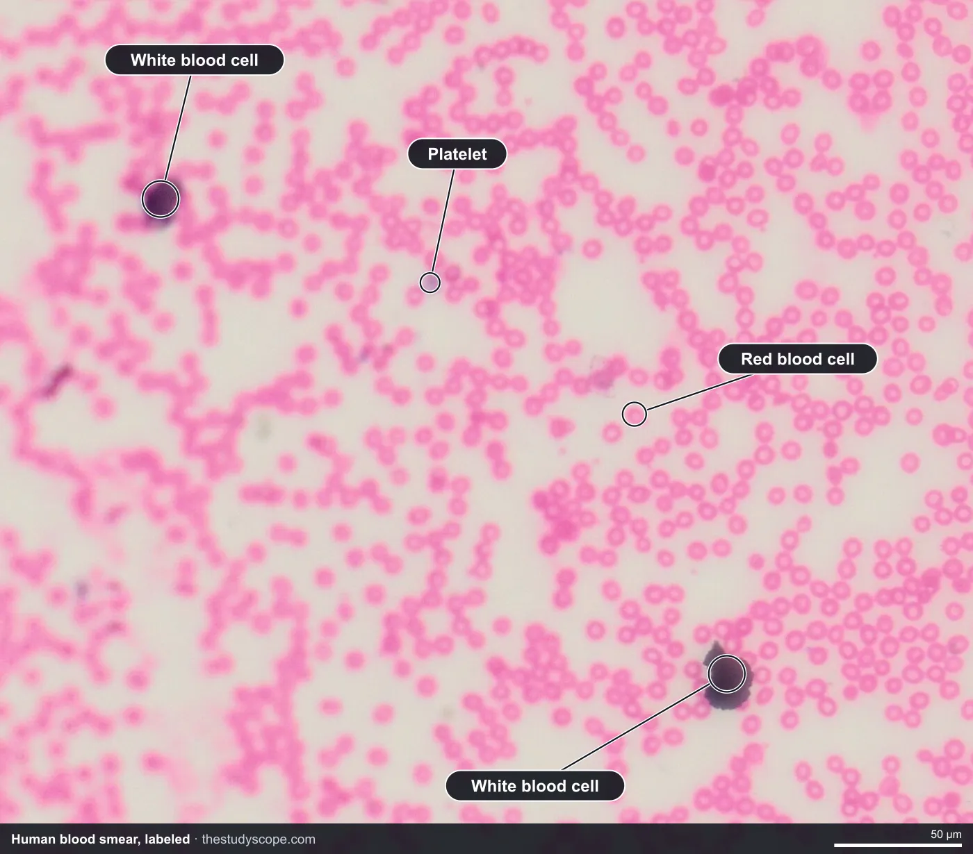
| Connective: blood | Round, pink red blood cells with no nucleus, and a few larger white blood cells with purple nuclei. | [Human blood smear](/catalog/slide/human-blood-smear) |
| Muscle | Long fibers with fine stripes and many nuclei at the edge. Cut across, they look like packed polygons in bundles. | [Skeletal muscle](/catalog/slide/skeletal-muscle-transverse-and-longitudinal-section) |
| Nervous | Not in the StudyScope catalog yet. | None |
The StudyScope catalog has no nervous tissue slide. In general, nervous tissue is made of neurons, which have a cell body and long thin extensions, packed among support cells, so it looks like cells with fine fibers running through them.
Labeled epithelial cells and blood
Epithelial tissue: cheek cells and the intestine lining
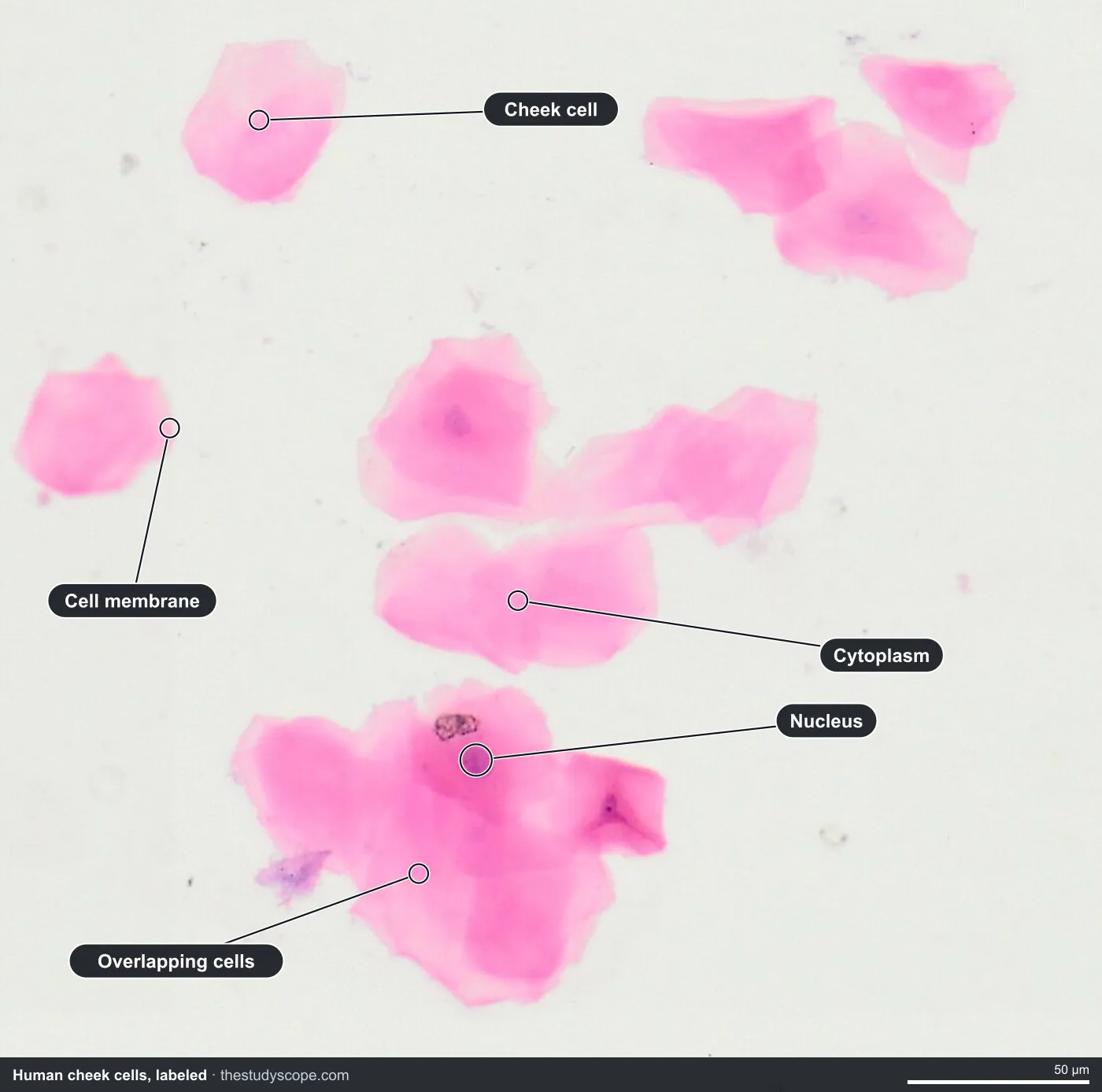
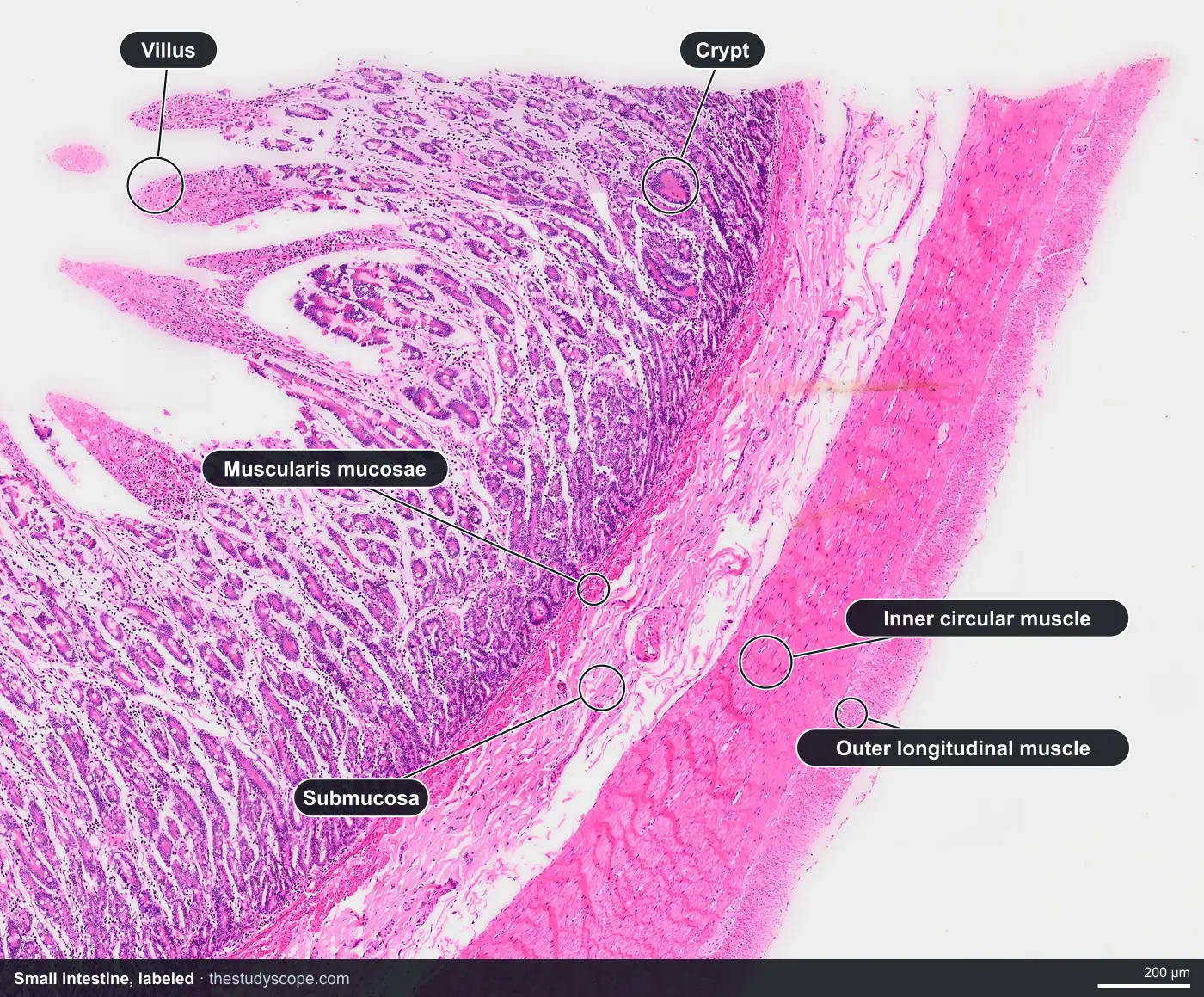
The cheek cell slide is a whole mount, meaning cells scraped from the inside of the mouth and spread on the slide, not a slice through a tissue. That makes it a good first look at single epithelial cells. The small intestine is a true cross section, so you see the epithelium in place as a lining.
- Cheek cells: thin, flat, many-sided cells, some alone and some overlapping in clumps, each with a small darker nucleus near the middle.
- Small intestine lining: finger-like villi reaching into the open center, covered by a single layer of tall cells with purple nuclei lined up near their base.
- Goblet cells: pale, cup-shaped cells among the lining cells that make mucus.
- What lies beneath: a layer of loose pink connective tissue (the submucosa), then two layers of smooth muscle.
How to identify hyaline cartilage under a microscope
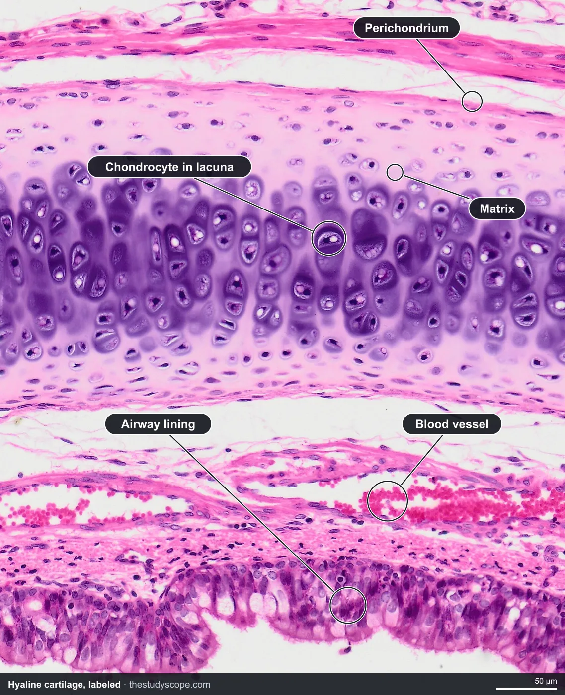
The hyaline cartilage slide is a section through the trachea (windpipe) stained with H&E. It cuts through a C-shaped ring of cartilage.
- Smooth matrix: a wide, glassy band stained light purple to bluish. It looks smooth, with no fibers or layers.
- Chondrocytes in lacunae: small cartilage cells sitting in little pockets scattered through the matrix, often in pairs or small groups, sometimes with a clear ring around the cell.
- No blood vessels: the cartilage itself has none.
- What surrounds it: on the airway side, a lining of tall cells with a fringe of cilia and pale mucus cells, then pink connective tissue between the lining and the cartilage.
A quick test for students: if the background is a smooth, glassy matrix with cells in pockets, think cartilage. If it is made of hard, layered rings with cells connected by channels, think bone.
How to identify skeletal muscle under a microscope
The skeletal muscle slide shows human muscle cut two ways, stained with iron hematoxylin. Because it is not H&E, the colors do not match the other slides, so rely on shape and pattern.
- Cut across (transverse): packed, many-sided shapes, each one a single long muscle fiber, grouped into bundles called fascicles and wrapped in connective tissue.
- Cut lengthwise (longitudinal): long fibers lying parallel, side by side.
- Striations: fine alternating light and dark bands across the long fibers. This is why skeletal muscle is also called striated muscle. They show best at high zoom.
- Nuclei at the edge: dark nuclei pressed against the outer edge of each fiber, not in the center, and many per fiber.
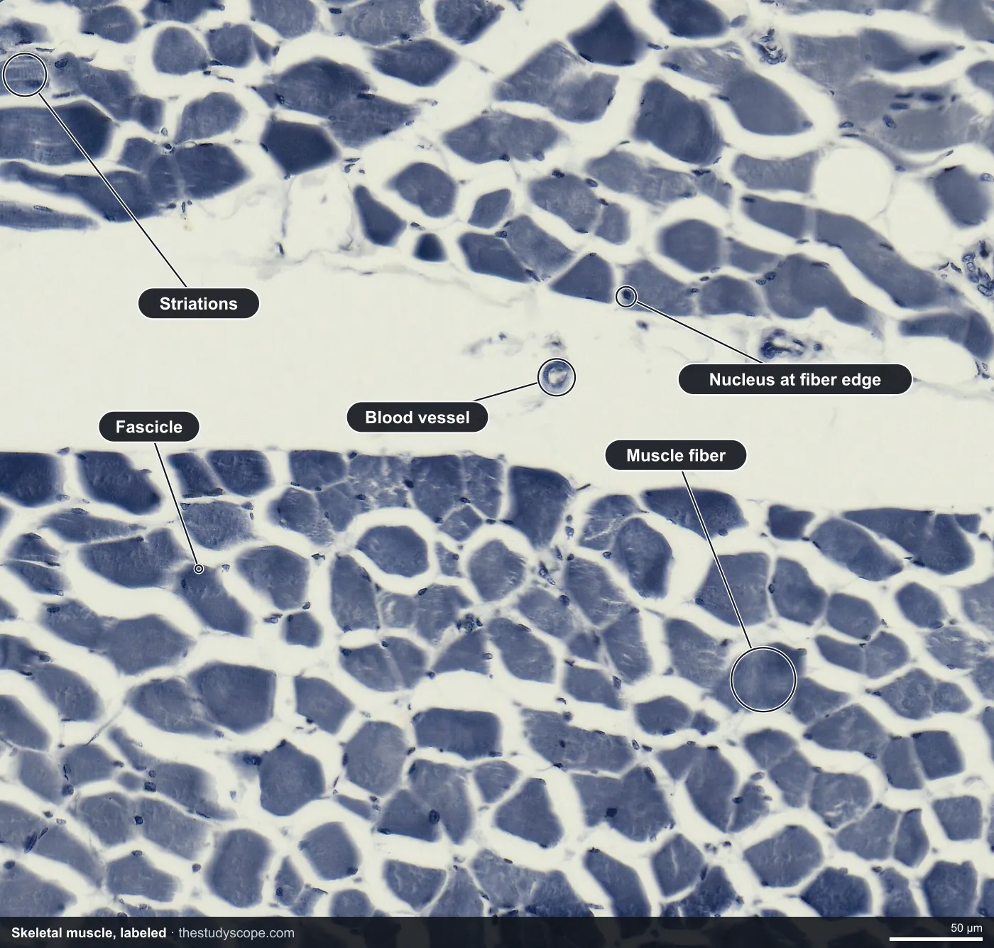
Smooth muscle has no stripes and is made of spindle-shaped cells with one central nucleus. You can see it as the muscle layers in the small intestine wall.
Putting it together: organ slides
Organs are made of several tissues in layers. These slides are good for practicing step one of the method, then naming the tissues you find.
- Small intestine: epithelium (the villi), connective tissue (submucosa), and smooth muscle, in order from the lumen outward.
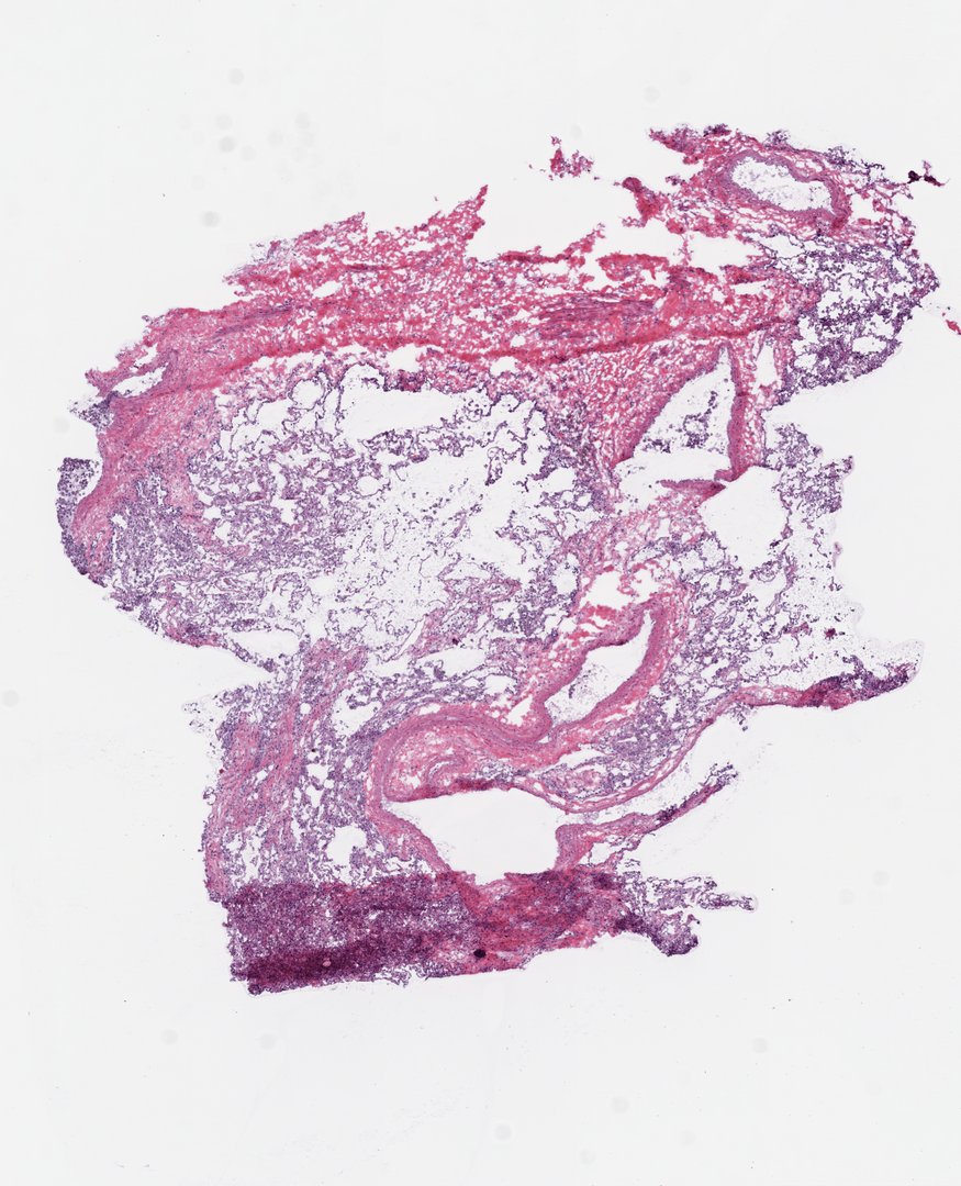
- Lung: a lacy network of tiny air sacs (alveoli) with very thin pink walls and purple nuclei. This is a frozen section, so some tears and folds come from the cutting, not the lung.
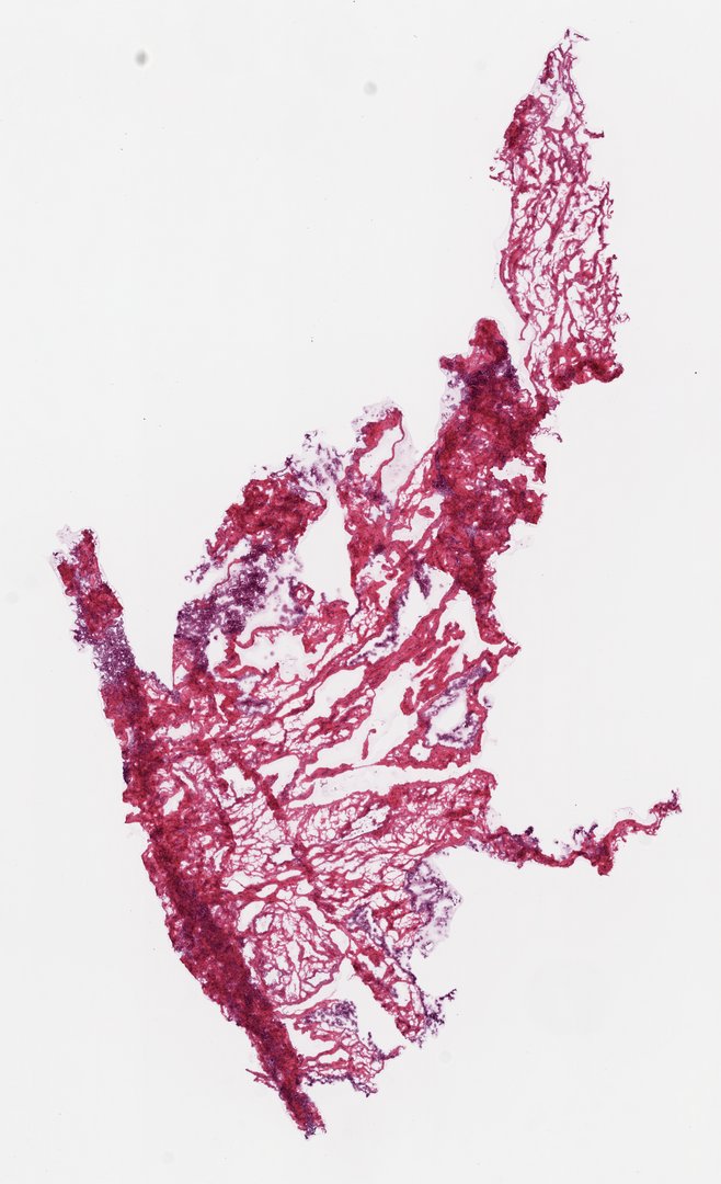
- Ovary: mostly stroma, a dense mix of spindle-shaped cells and pink collagen fibers, with blood vessels running through. Also a frozen section.
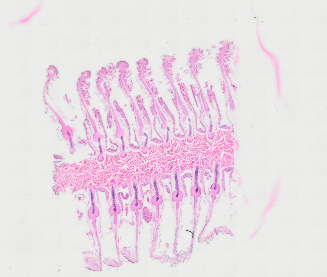
- Fish gill: rows of long filaments off a supporting arch, with thin plates (lamellae) full of red blood cells, and a cartilage rod inside each filament.
- Frog skin: an epidermis of several rows of cells with purple nuclei, over a dermis of pink connective tissue with round glands.
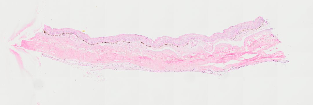
Reading histology slides online with a class
- Present a slide live and have students follow on their own screens with a room code. No student accounts are needed.
- Begin at the lowest zoom and ask students to sketch the layout and name the layers before you zoom in.
- Zoom to the cells and ask students to decide the tissue type from the evidence: packed cells, scattered cells in matrix, or long fibers.
- Use the ruler to measure a nucleus or a fiber. Every slide is calibrated, so the numbers are real micrometers.
- Students can draw boxes around what they found and save a stamped snapshot of their view for their notebooks.
One honest note: a bench microscope with a drawer of glass slides shows you the same kind of tissue, but a class shares one view at a time through the eyepiece. Online, the whole room looks at the same spot together.
Browse the full slide catalog without an account, or open hyaline cartilage, skeletal muscle, or the small intestine to start practicing the method. Deep zoom on these slides needs a teacher seat; see pricing.
Questions and answers
How do you read a histology slide?
Start at the lowest zoom and look at the overall layout: the edges, any open spaces, and the layers. Then check the stain, since in H&E nuclei are purple and most other tissue is pink. Finally zoom in to see the shape and arrangement of the cells and where their nuclei sit.
What are the 4 types of tissue under a microscope?
Epithelial tissue (packed cells forming linings and coverings), connective tissue (cells in a lot of material between them, including cartilage and blood), muscle tissue (long fibers), and nervous tissue (neurons and support cells). StudyScope has slides for epithelial, connective, and muscle tissue, but not nervous tissue.
How do you identify hyaline cartilage under a microscope?
Look for a smooth, glassy, light purple to bluish matrix with small cells called chondrocytes sitting in little pockets called lacunae, often in pairs or small groups. Hyaline cartilage has no blood vessels. In the trachea slide it forms a C-shaped ring.
How do you tell skeletal muscle from other tissues under a microscope?
Skeletal muscle is made of long, parallel fibers with fine light and dark stripes (striations) and many nuclei at the edge of each fiber. Cut across, the fibers look like packed polygons grouped into bundles.
Why are histology slides pink and purple?
Many are stained with H&E. Hematoxylin colors nuclei purple and eosin colors most other tissue pink. Other stains give other colors, so check the stain listed for each slide.
Where can I look at histology slides online?
StudyScope is an online virtual microscope with real prepared slides, including small intestine, lung, skeletal muscle, hyaline cartilage, and blood. You can browse the catalog and previews without an account. It runs in any modern browser, including Chromebooks and iPads.
More guides
- Monocot vs dicot stems compared
- Frog, human, chicken and fish blood compared
- Virtual microscope, simulator or digital microscope?
- Using a microscope on a Chromebook
- How to identify the stages of mitosis in an onion root tip
- Cheek cells vs onion cells
- Leaf layers and stomata guide
- Pond water organisms identification guide
- How to measure cell size
- Lily, pine and luffa pollen compared
- Insect mouthparts compared
- Worm body cavities compared