Short answer: To identify pond water organisms, start with the shape and size, then look for one key feature: a slipper-shaped cell is a paramecium, a spindle-shaped cell is a euglena, green threads with a spiral ribbon inside are spirogyra, bead-like chains in jelly are nostoc, and anything with a shell, antennae or a tail fork is a small animal such as daphnia, cyclops or a mosquito larva. The slides on this page are real prepared, stained or mounted slides of each organism, not a live pond-water sample. Nothing on them moves, so they teach what each organism looks like, and a live wet mount shows the swimming that a fixed slide cannot.
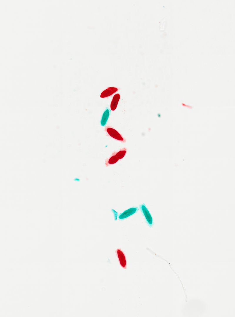 Paramecium (Whole Mount)ParameciumVarious
 Euglena (Whole Mount)EuglenaVarious
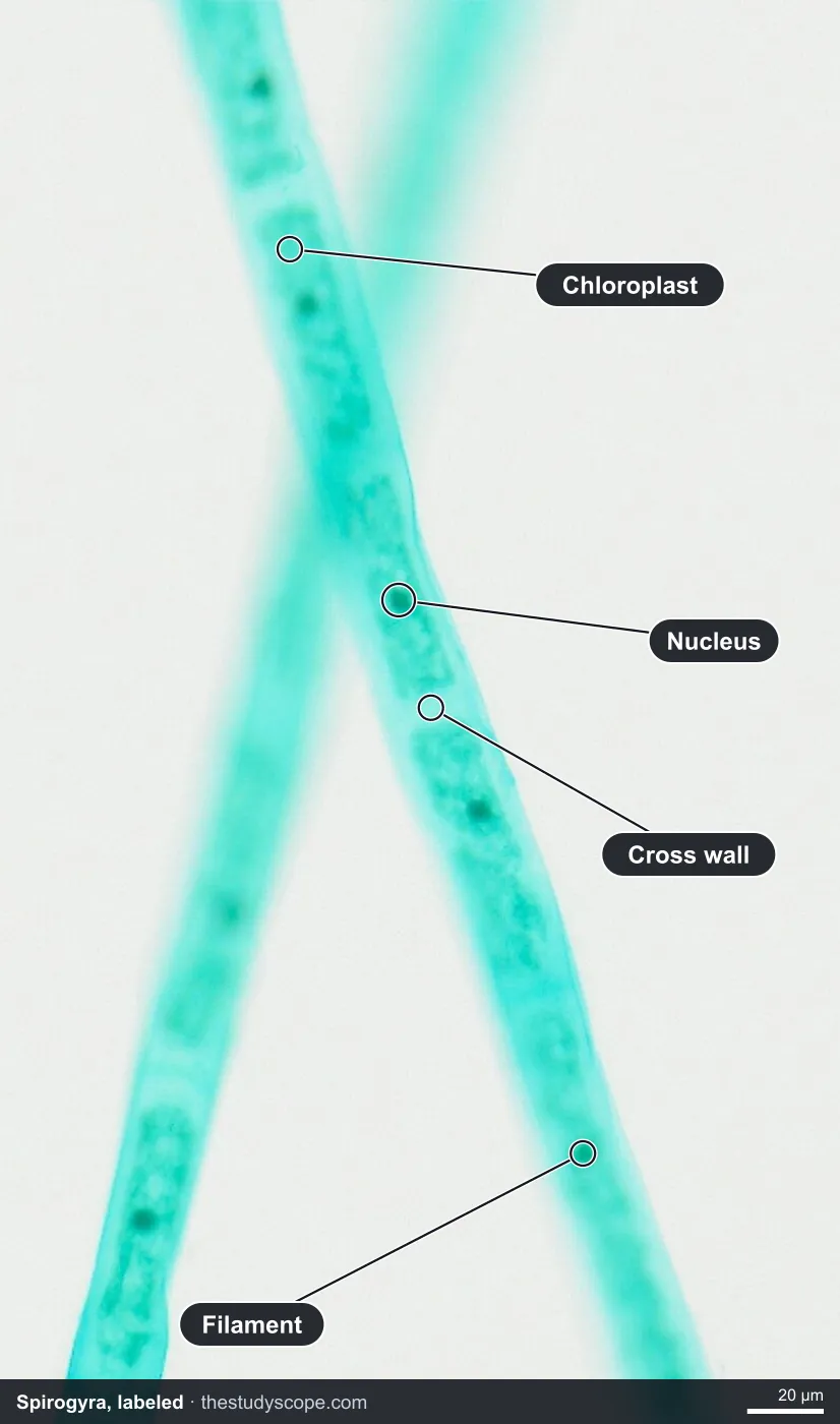
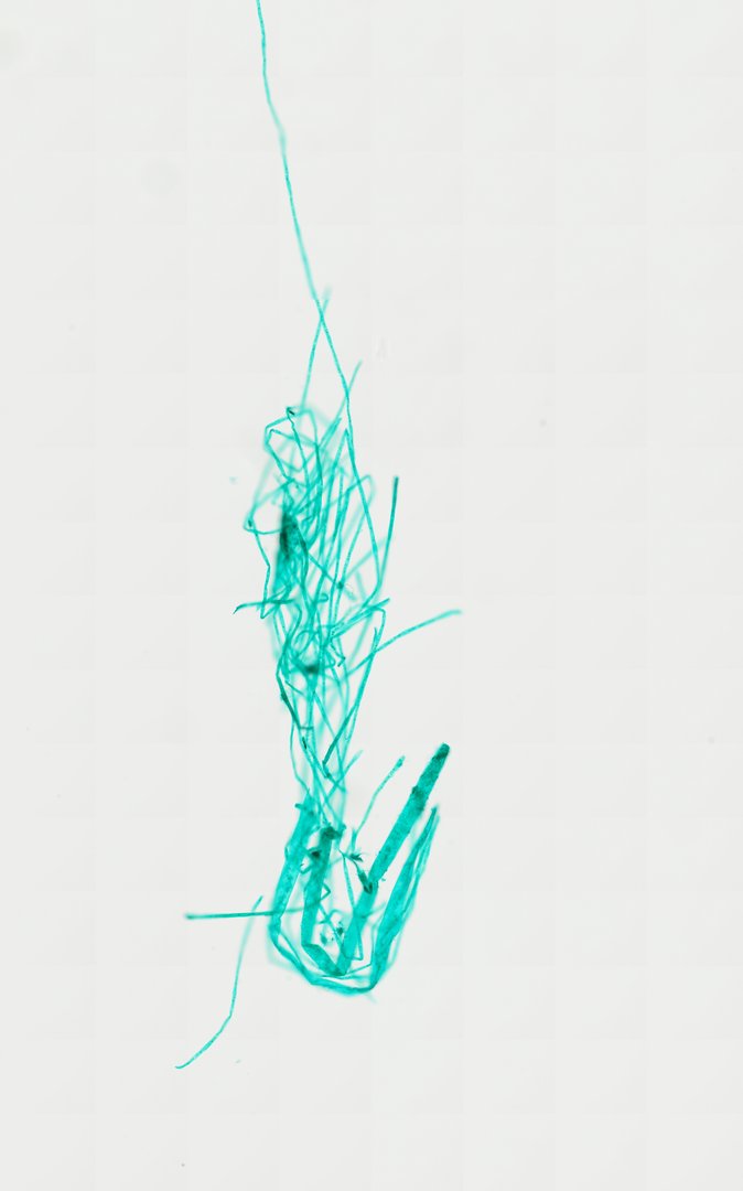 Spirogyra (Whole Mount)SpirogyraFast Green
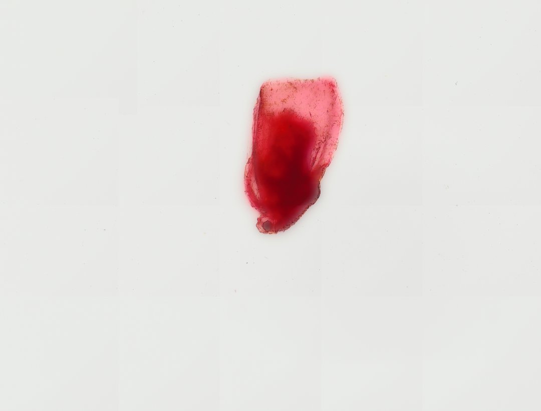 Daphnia (Whole Mount)DaphniaCarmine
Prepared slides vs a live pond-water sample
A drop of real pond water under a bench microscope is alive. Cells swim, cilia and flagella beat, and a daphnia's heart pulses. A prepared slide is a different tool. The organism was fixed, stained or mounted by a professional, so it holds still, the colors are the stain, and details like cilia and flagella are often faint. What you gain is a clean, well-prepared example of each organism that you can study at your own pace, which is hard to get from a random drop of pond water where everything is moving and mixed together.
Pond water microorganisms identification chart
| Organism | Group | What you notice first | Key feature to look for on our slide |
|---|---|---|---|
| Paramecium | Protist (ciliate) | Many slipper-shaped cells, about 0.2 millimeters long, stained red or teal | Rounded end and narrower end, the oral groove, food vacuoles; cilia at the cell edge |
| Euglena | Protist (flagellate) | Many long spindle-shaped cells, often around 50 micrometers | Pointed ends, a flexible pellicle, a nucleus; a thin flagellum on a few cells |
| Chlamydomonas | Alga (single-celled green alga) | Thousands of tiny oval cells, about 10 micrometers each, stained with Fast Green | Cup-shaped chloroplast and pyrenoid at high zoom; the two flagella are very thin |
| Spirogyra | Alga (filamentous green alga) | Long unbranched green threads crossing the slide | Ribbon-shaped chloroplast winding in a spiral, pyrenoids, a nucleus in the middle of the cell |
| Nostoc | Cyanobacterium | A long brown band of colony in thick jelly | Bead-like chains and heterocysts, though the dense jelly makes single cells hard to pick out |
| Daphnia (water flea) | Crustacean | A whole oval animal in a clear shell, stained red with carmine | Carapace edge, a single compound eye, large branched antennae |
| Cyclops | Crustacean (copepod) | A teardrop-shaped body, one to two millimeters, with a narrow tail | Forked tail with long bristles, long antennules, swimming legs, egg sacs on some |
| Mosquito larva (Culex) | Insect larva | A long, unstained, segmented body | Head with dark eye spots, wide thorax, hair tufts, and the breathing siphon near the tail |
Paramecium and spirogyra under the microscope, labeled
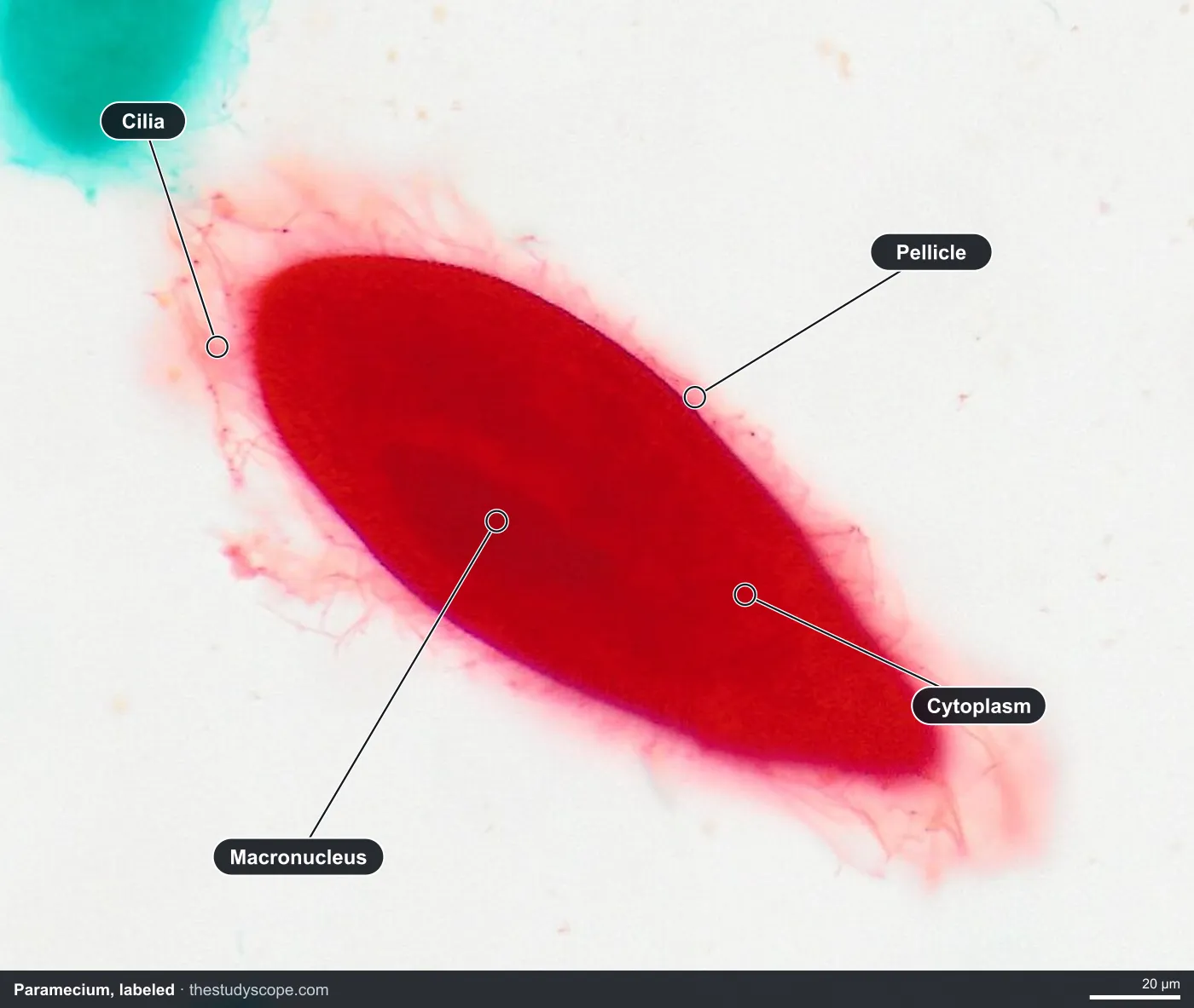
Protists under the microscope: paramecium, euglena and chlamydomonas
A protist is a mostly single-celled organism that is not a plant, an animal or a fungus. These three are the classic set for telling different protists apart under the microscope.
Paramecium
- Slipper shape: an oval cell, rounded at one end and narrower at the other.
- Oral groove: a dent along one side where food is swept in.
- Macronucleus and micronucleus: a large dark nucleus near the middle, and a much smaller one beside it that can be hard to see.
- Food vacuoles: small round bubbles inside the cell, at high zoom.
- Cilia: very fine hairs covering the cell. They are often hard to see on a stained slide, so look at the edges.
Euglena
- Spindle shape: long cells, wide in the middle and tapered at both ends.
- Pellicle: a flexible outer covering that lets the cell bend and change shape.
- Nucleus and chloroplasts: visible inside the cell at high zoom.
- Flagellum: a thin thread trailing from one end. It shows on only a few cells here, because it is fine and easy to lose on a prepared slide.
Euglena is a good puzzle for a class. It can make food like a plant but swims and takes in food like an animal, so it does not fit neatly into either group.
Chlamydomonas
- Tiny oval cells: thousands of them, each only about 10 micrometers long, so this is the smallest organism in the set.
- Cup-shaped chloroplast: one large chloroplast fills much of each cell.
- Pyrenoid: a small round body inside the chloroplast, where starch is made.
- Flagella and eyespot: two thin whip-like threads at one end and a light-sensing spot. Both are hard to see after staining.
Algae and cyanobacteria under the microscope: spirogyra and nostoc
Spirogyra is a green alga that grows as unbranched threads. Each thread is a chain of cylinder-shaped cells, and each cell holds a ribbon-shaped chloroplast that winds in a spiral. Pyrenoids are spaced along the ribbon, and the nucleus sits near the middle of the cell. It is the easiest pond organism to identify on sight.
Nostoc looks different because it is not an alga. It is a cyanobacterium, a kind of bacteria that makes its own food by photosynthesis, which is why cyanobacteria under the microscope were once called blue-green algae. Its cells have no nucleus. Nostoc grows as bead-like chains packed in a thick jelly. Some cells in the chain, called heterocysts, capture nitrogen from the air. On our slide the colony shows as a long brown band, and the jelly is dense enough that single cells are hard to pick out, so use it to show the colony form and the jelly, and use high zoom to hunt for the beaded chains.
Small pond animals: daphnia, cyclops and mosquito larva
- Daphnia (Whole Mount)DaphniaCarmine
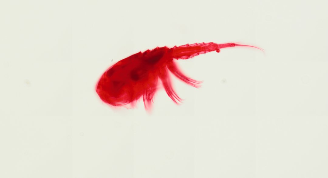 Cyclops (Whole Mount)Cyclops sp.Safranin
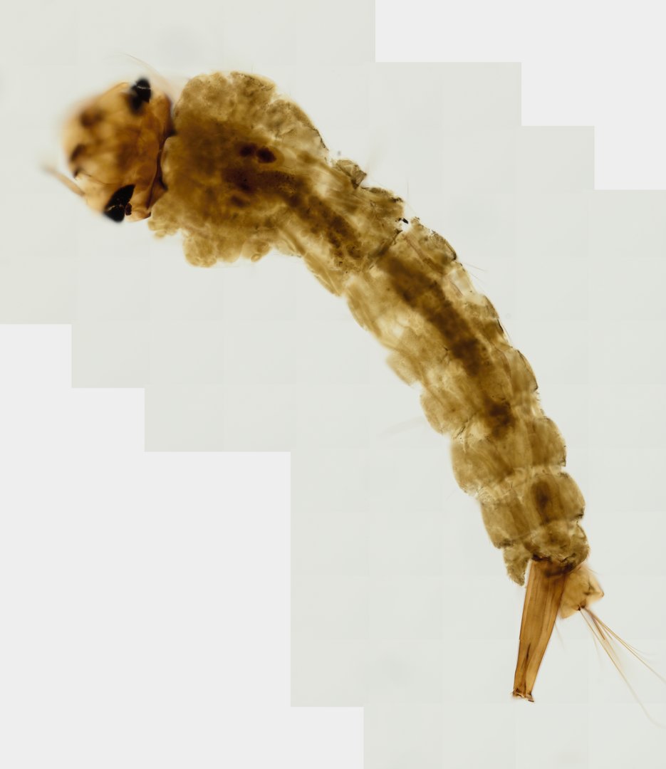 Mosquito Larva (Whole Mount)Culex sp.Unstained
- Daphnia: the water flea, a tiny freshwater crustacean. Look for the pale pink carapace edge, the single dark compound eye, and the large branched antennae it rows with. The organs are packed tightly under the stain, so use it for the outline, not for picking out each organ.
- Cyclops: a copepod, another crustacean, stained red with safranin. The front body is wide and rounded, then narrows into a segmented tail that ends in a fork with long bristles. Several pairs of swimming legs hang from underneath, and females may carry a pair of egg sacs.
- Mosquito larva: a Culex larva, left unstained so its natural color shows. Find the head with dark eye spots, the wide thorax, the segmented abdomen with hair tufts, and the long breathing siphon near the tail that it pushes up through the water surface to get air.
Telling the three apart is easy: daphnia has a shell and a big oval body, cyclops has a narrow tail with two bristled forks, and the mosquito larva is a long, segmented insect body with a breathing tube.
Other pond life on the catalog
Some pond and still-water animals are only available as sections, not whole mounts, so they show inner structure instead of the whole organism. The planaria cross section shows a freshwater flatworm with no body cavity and a branched gut. The leech cross section shows the thick muscle and tissue of Hirudo. Bacteria are also in the catalog, on the three types of bacteria smear, which shows the three basic shapes (round cocci, rod-shaped bacilli and corkscrew spirilla), but it is a prepared smear and not a pond sample.
Using the slides in a pond water lab
- Before the lab, present the identification chart and a few slides live, so students have a picture of each organism in their heads. Students join with a room code, with no accounts, and follow on their own screens.
- Run the real pond-water lab with wet mounts and bench microscopes, where students see the swimming, the cilia and the flagella that a fixed slide cannot show.
- After the lab, have students match what they sketched to a prepared slide. A fixed, stained organism is much easier to name, and it settles arguments about what that blob was.
- Use the ruler to measure one cell on each slide. Every slide is calibrated, so a paramecium and a chlamydomonas can be compared in real micrometers.
- Have students draw a box around the key feature on one slide and save a stamped snapshot of their view as evidence for their identification.
Teachers can use these slides to teach identification either before or after a real pond-water lab. Open a slide from the set above, or browse the whole catalog without an account.
Questions and answers
How do you identify organisms in pond water under a microscope?
Check size and shape first, then look for one key feature. Paramecium is a slipper-shaped cell, euglena is spindle-shaped, spirogyra is a green thread with a spiral chloroplast, daphnia is a small animal in a clear shell, and cyclops has a forked tail with long bristles. Movement is a big clue in a live sample, which is why a chart of still, prepared slides works best alongside a real pond-water lab.
What does paramecium look like under a microscope?
Paramecium is a slipper-shaped single cell, rounded at one end and narrower at the other, about 0.2 millimeters long. At higher zoom you can find the oral groove, food vacuoles, and a large macronucleus. Its cilia are very fine and can be hard to see on a stained slide.
What is the difference between euglena and chlamydomonas?
Both are single cells that swim with flagella and can photosynthesize. Euglena is a longer, spindle-shaped cell, often around 50 micrometers, with a flexible pellicle. Chlamydomonas is a much smaller oval cell, about 10 micrometers, with one cup-shaped chloroplast and two flagella.
Are cyanobacteria under the microscope bacteria or algae?
Cyanobacteria are bacteria. They photosynthesize like algae and were once called blue-green algae, but their cells have no nucleus. Nostoc is a classic example, with bead-like chains of cells packed in jelly and special nitrogen-capturing cells called heterocysts.
Can you see paramecium moving on a prepared slide?
No. A prepared slide is fixed and stained, so nothing moves, and cilia and flagella are often faint. To watch paramecium swim, you need a live wet mount on a bench microscope. A prepared slide is better for studying the shape and structure of the cell.
Is this a live pond water sample?
No. Every slide here is a professionally prepared slide of one organism, stained or mounted and then scanned. They are meant to teach identification, and they pair well with a real pond water lab with a live sample.
More guides
- Monocot vs dicot stems compared
- Frog, human, chicken and fish blood compared
- Virtual microscope, simulator or digital microscope?
- Using a microscope on a Chromebook
- How to identify the stages of mitosis in an onion root tip
- Cheek cells vs onion cells
- Leaf layers and stomata guide
- How to identify tissue types
- How to measure cell size
- Lily, pine and luffa pollen compared
- Insect mouthparts compared
- Worm body cavities compared