Short answer: Lung tissue looks like lace under the microscope because most of it is air. The open spaces are alveoli, tiny air sacs, and the pink lines between them are walls so thin that oxygen crosses them by diffusion into the blood in their capillaries. Scattered through the lace are blood vessels with thick pink walls and, in most adult lungs, black specks of carbon. Gills solve the same problem in water. A fish gill packs thin plates called lamellae along rows of filaments, and an oyster gill uses rows of filaments to breathe and to catch food.
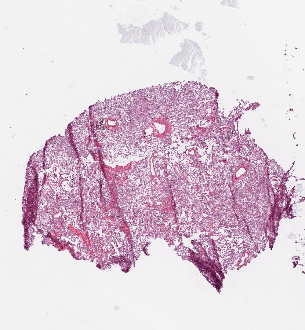 Lung (top section) Slice 3Homo sapiensH&E
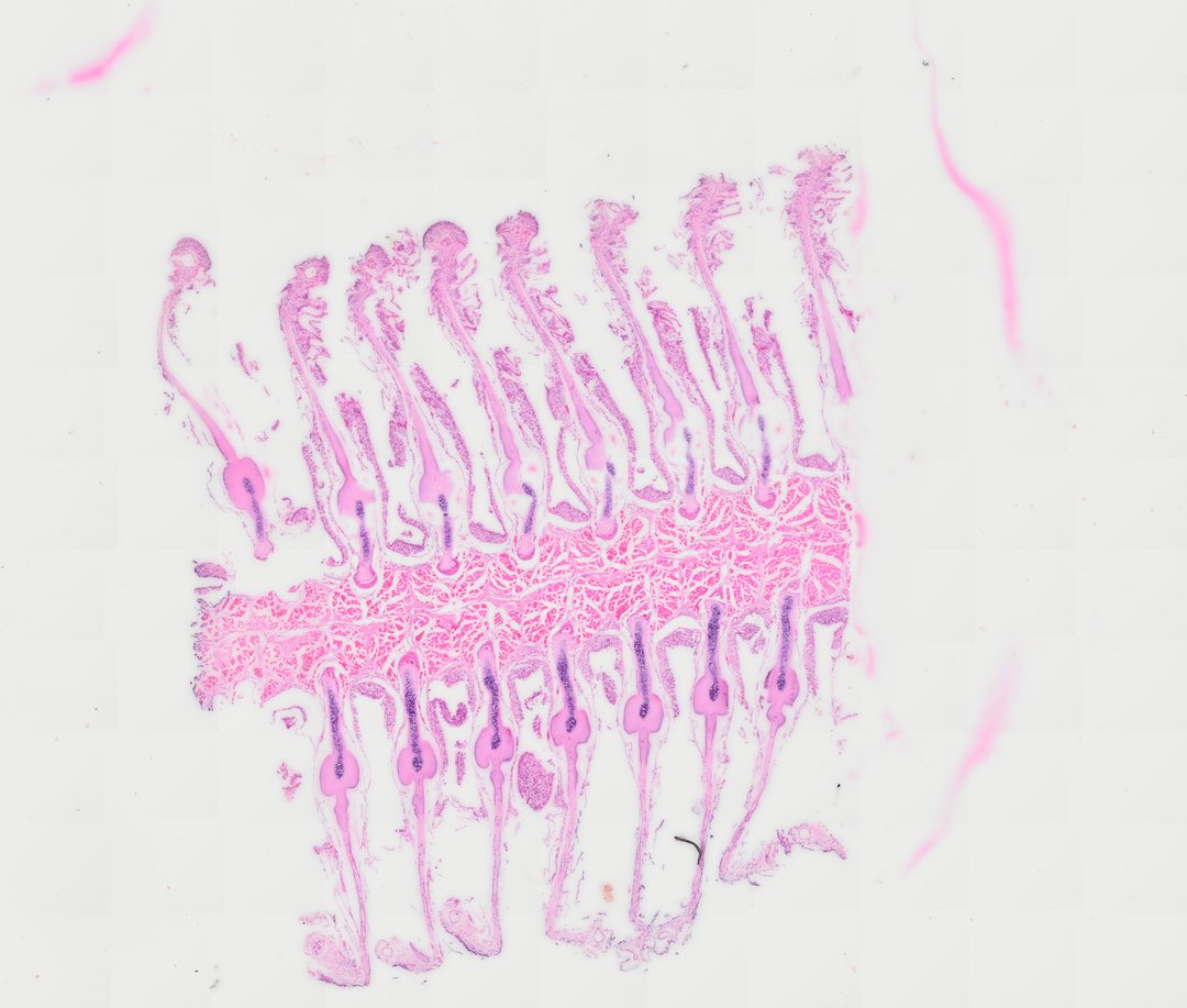 Fish Gill (Cross Section)ActinopterygiiH&E
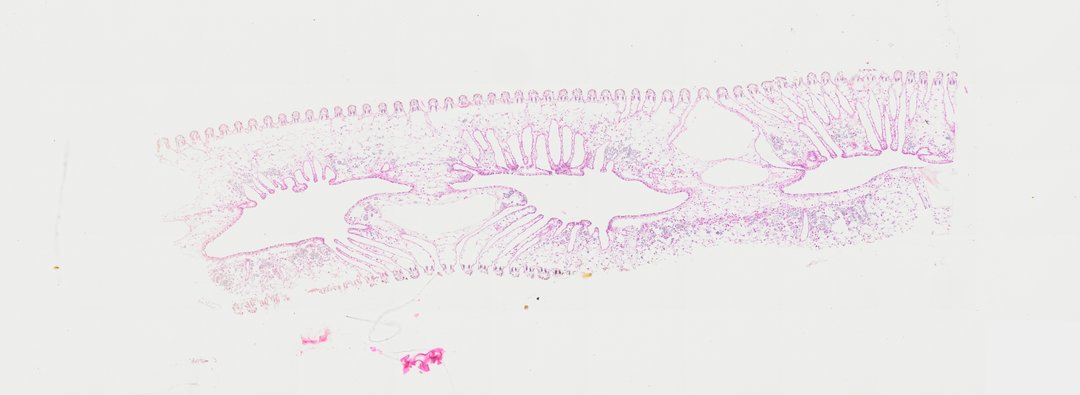 Oyster Gill (Cross Section)Crassostrea virginicaH&E
What lung tissue looks like under the microscope
At low zoom a lung section is a pink and purple net with more white space than tissue. Each hole in the net is an alveolus, or part of one. In an adult lung an alveolus is about 0.2 millimeters across, and there are hundreds of millions of them, enough wall to cover roughly 70 square meters if you spread it flat. Zoom in on a single wall and it is only one or two cells thick, with flat purple nuclei. That thinness is the whole point: diffusion is fast over a short distance and slow over a long one.
Lung tissue labeled: what to look for
Lung Slice 3 shows the most in one place, so start there.
- Alveoli: the open, empty-looking spaces that make the lace.
- Alveolar walls (septa): thin pink strands between the spaces, dotted with dark purple nuclei.
- Blood vessels: round or oval rings with a thick pink wall that looks wavy at high zoom. Several sit in the upper half of the section, and they stand out from the lace at low zoom.
- Carbon pigment: black clumps, often right next to the vessels. This is soot and dust breathed in over a lifetime. Most adult lungs have some.
- Collapsed edges: dark, crowded bands along the sides and bottom, where the air sacs have closed and the walls are squeezed together.
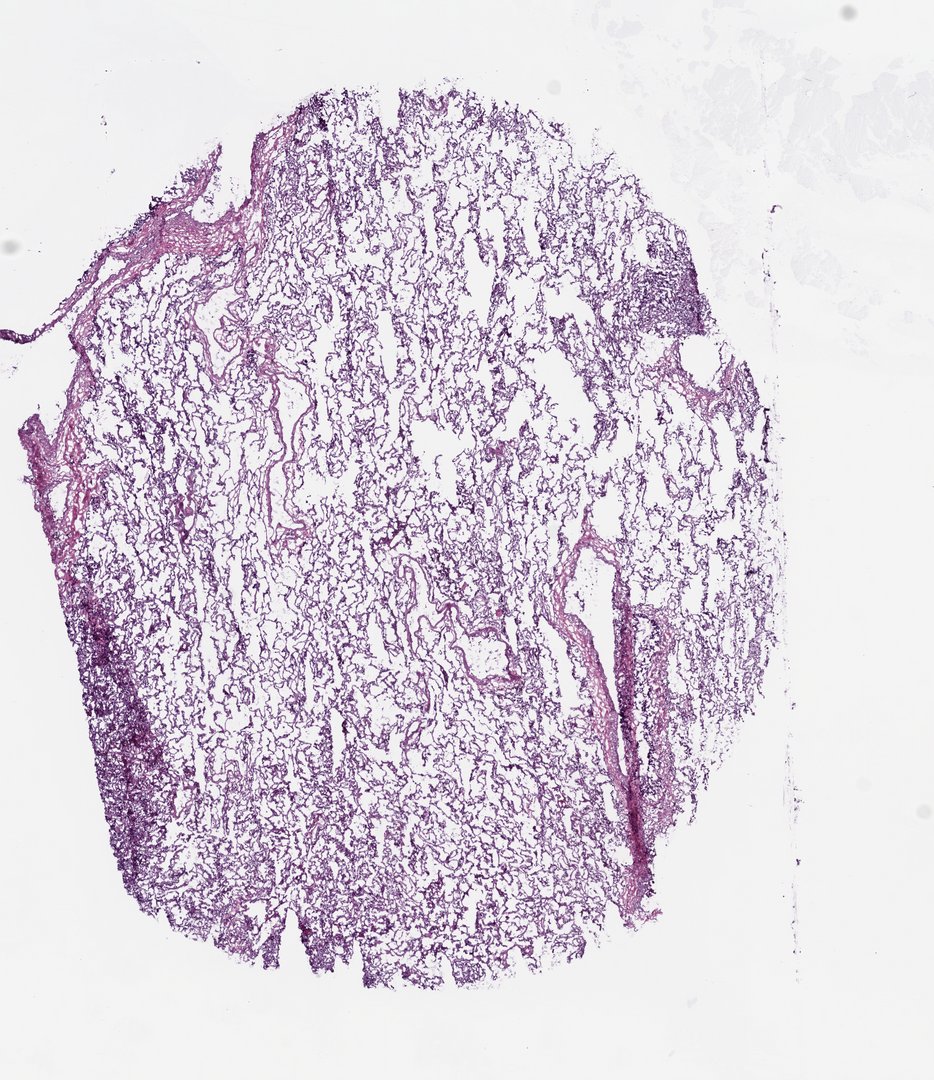
Lung Slice 4 is the cleanest lace in the series: open alveoli edge to edge, with a dense collapsed strip down the left side. Its stain runs more purple than pink, so judge by shape, not color. It is the better slide for measuring alveoli.
These are frozen sections
All six lung slides in the catalog are frozen sections of normal lung taken next to a tumor. Freezing and cutting soft, air-filled tissue leaves marks: long folds where the slice creased, tears and holes, and collapsed edges. They are worth pointing out, because students should learn to tell the specimen from the way it was prepared. A creased fold runs in a straight line across many alveoli at once; a real wall curves around a single air space.
- Slice 1: lots of bright pink and red where blood pooled, a large vessel near the top, and a thick, dark collapsed band along the bottom.
- Slice 2: open lace with bigger tears and holes, and a few black carbon specks.
- Bottom Slice 1: the most torn of the six, in loose pieces. Use it to talk about artifacts, not to learn the structure.
- Bottom Slice 2: lace crossed by long diagonal fold lines, with a collapsed band along one edge.
Fish gill under the microscope
The fish gill is cut through part of a gill arch. A thick band of muscle runs across the middle, and long gill filaments stick out from it in two rows, like the teeth of two combs set back to back.
- Gill filaments: the long fingers on both sides of the central band.
- Cartilage rods: a stiff pink rod down the middle of each filament, packed with purple cartilage cells near its base. It holds the filament out in the water.
- Lamellae: thin flaps of tissue along the sides of each filament, only a few cells thick. Some have lifted or torn away in this section.
- Blood cells: rows of small cells inside the lamellae. Fish red blood cells keep their nucleus, so each one shows a purple dot.
- Muscle: the pink blocks in the central band, cut across.
Oyster gill under the microscope
The oyster gill looks very different. An oyster breathes with its gill and also feeds with it: in a living oyster, tiny hairs called cilia beat to pull water through, and food particles get trapped in mucus and moved toward the mouth.
- Gill filaments: a long row of small rounded bumps along the outer edge of the section, evenly spaced like beads.
- Supporting rods: a pair of thin rods that stain purple inside each filament.
- Water tubes: large open spaces inside the gill, where water flows after passing between the filaments.
- Tissue bridges: thin pink walls that cross the water tubes and join the two sides of the gill.
- Blood cells: round cells scattered through the loose tissue.
The cilia themselves are too fine to pick out on this scan, so treat them as the reason for the filaments, not something to find.
Gills vs lungs at a glance
| Feature | Human lung | Fish gill | Oyster gill |
|---|---|---|---|
| Breathes | Air | Water | Water |
| Exchange surface | Walls of the alveoli | Lamellae along each filament | Filaments along the gill surface |
| How the air or water moves | In and out the same way, by breathing | One way, in through the mouth, over the gills, out under the gill cover | One way, pulled through by beating cilia |
| What holds it open | Elastic tissue, with the chest wall pulling the lung open | A cartilage rod in each filament | A pair of thin rods in each filament |
| Extra job | None | None | Catches food (filter feeding) |
| Look for on the slide | Lace of alveoli, thick-walled vessels, black carbon | Two rows of filaments, cartilage rods, thin lamellae | Beaded row of filaments, large water tubes |
Why lungs and gills share a design
Oxygen moves into blood by diffusion, and diffusion only works well across short distances and large areas. So every one of these surfaces is thin, folded to fit a lot of area into a small space, and has blood running right under it. What changes is the medium. Water holds far less oxygen than air, which is why fish push water one way over the lamellae while blood flows through them in the opposite direction. That countercurrent flow lets the blood keep picking up oxygen along the whole lamella. Air is thin and light enough to pump in and out of dead-end sacs. Pull a fish out of water and the opposite problem appears: without water to hold them apart, the lamellae stick together and most of the surface is lost.
Teaching it with StudyScope
- Measure both surfaces. Every slide is calibrated, so the ruler reads real micrometers. Have students measure an alveolus on Slice 4 and a lamella on the fish gill, then compare.
- Present it live. Students join with a room code on their own screens, with no student accounts, and follow your view as you move from lung to gill.
- Name the artifacts. Ask students to find one fold and one real alveolar wall on the same lung slide and explain how they told them apart.
Want it ready to teach? The premade lesson Gas Exchange: Lungs and Gills is about 35 minutes. Students compare human alveoli, fish gill lamellae and an oyster gill, and you can present it live while every student screen follows. Browse the catalog without an account.
Questions and answers
What does lung tissue look like under a microscope?
Lung tissue looks like lace or a sponge. Most of it is open air sacs called alveoli, separated by very thin walls. With H&E stain the walls are pink with dark purple nuclei, blood vessels have thick pink walls, and black specks of carbon are common in adult lungs.
What are alveoli and why are their walls so thin?
Alveoli are the tiny air sacs at the ends of the airways, about 0.2 millimeters across in an adult. Oxygen passes from the air into the blood by diffusion, which only works well over short distances, so the wall between air and blood is kept to one or two thin cells.
What are the black spots in lung tissue?
They are carbon pigment: soot, smoke and dust breathed in over the years and stored in the lung, often near blood vessels. Most adult lungs have some, smokers and city dwellers more.
What does a fish gill look like under a microscope?
A fish gill has rows of long filaments coming off a supporting arch, each held out by a cartilage rod. Thin plates called lamellae line both sides of each filament and are full of blood cells, which in fish have a nucleus.
What is the difference between gills and lungs?
Both are thin, folded surfaces with blood just underneath, built for diffusion. Lungs take in air and move it in and out of dead-end air sacs. Gills take in water and move it one way over the lamellae, often against the direction of blood flow, which helps because water holds much less oxygen than air.
Why can't fish breathe out of water?
Water holds the thin lamellae apart. In air they collapse and stick together, so most of the gill surface is lost, and the fish cannot take in enough oxygen even though air has more of it.
More guides
- Monocot vs dicot stems compared
- Frog, human, chicken and fish blood compared
- Virtual microscope, simulator or digital microscope?
- Using a microscope on a Chromebook
- How to identify the stages of mitosis in an onion root tip
- Cheek cells vs onion cells
- Leaf layers and stomata guide
- Pond water organisms identification guide
- How to identify tissue types
- How to measure cell size
- Lily, pine and luffa pollen compared
- Insect mouthparts compared
- Worm body cavities compared
- Insect wings compared