Orange Peel Cross Section Under the Microscope
Orange Peel (Cross Section), Citrus sinensis
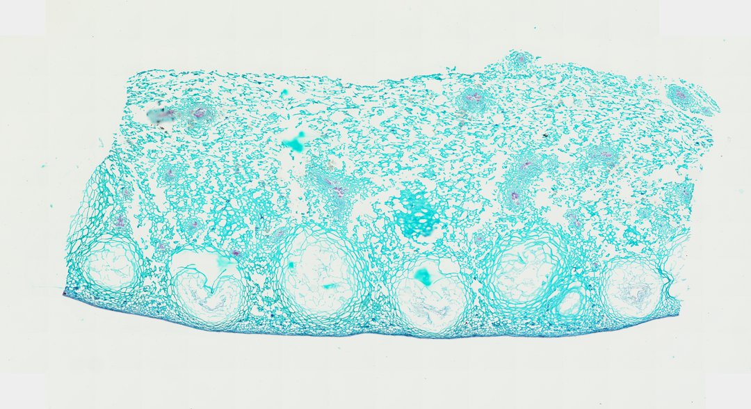
This is a cross section of orange peel stained with safranin and fast green. It shows the colorful outer layer with oil glands and the white, spongy layer underneath.
This cross section of an orange peel reveals the structural zonation characteristic of citrus rinds. The outer flavedo (exocarp) consists of compact epidermal cells covered by a thick cuticle, interspersed with prominent, spherical oil glands (schizolysigenous cavities) bounded by specialized epithelial secretory cells that synthesize essential oils. Deep to the flavedo lies the expansive albedo (mesocarp), composed of a porous, spongy meshwork of branched parenchyma cells (aerenchyma) interspersed with large intercellular air spaces. Small vascular bundles containing lignified xylem vessels and phloem elements are distributed throughout the mesocarp matrix. Differential staining emphasizes cell wall composition, highlighting cellulosic parenchyma walls alongside specialized epidermal and secretory boundaries.
What to look for
- Flavedo: The outer layer of the peel, made of small, compact cells.
- Oil glands: Large round cavities in the outer layer where citrus oils are stored.
- Albedo: The spongy inner layer of loosely connected cells with large air spaces.
- Vascular bundles: Small veins in the albedo, with xylem stained red.
At each magnification
Low power (about 4x)
A band of tissue with large round holes near the outer edge.
Medium power (about 10x)
The oil glands in the outer layer and the loose meshwork of the inner layer.
High power (about 40x)
The cells lining the oil glands and the branched cells of the spongy layer.
Questions and answers
What does orange peel look like under a microscope?
Orange peel has a thin outer layer (the flavedo) with large, round oil glands, and a thicker white layer (the albedo) made of loose, spongy cells with lots of air spaces.
Where does the smell of an orange peel come from?
The smell comes from oils stored in the round oil glands in the outer peel. When you bend or scratch the peel, the glands break and release the oil.
- Category
- Botany & Plants
- Scan magnification
- 20x
- Stain
- Safranin / Fast Green
- Tags
- orange peel, Citrus sinensis, citrus rind, flavedo, albedo, exocarp
Viewing this slide in class
This is a scan of a real prepared slide, not a simulation or a drawing. On StudyScope you can pan from the whole slide down to individual structures, always in focus, and measure them in real micrometers with the calibrated ruler. Present it live and every student screen follows your view and annotations. Students join with a room code on Chromebooks, iPads, or any browser, with no accounts.