Cotton Leaf Cross Section Under the Microscope
Cotton Leaf (Cross Section), Gossypium hirsutum
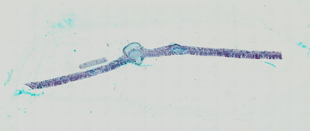
This is a cross section of a cotton leaf stained with safranin and fast green. It shows the classic layers of a dicot leaf, from the upper skin through the food-making cells to the vein in the middle.
A cross section of a cotton leaf (Gossypium hirsutum C.S.) double-stained with Safranin and Fast Green, highlighting the classic dorsiventral tissue organization of a dicotyledonous leaf. A prominent midrib protrudes abaxially, housing a well-developed crescent-shaped primary vascular bundle composed of lignified xylem vessels oriented toward the adaxial surface and phloem tissue positioned abaxially. The upper and lower surfaces are bounded by single-layered epidermal cells. Beneath the adaxial epidermis, a densely packed layer of elongated palisade mesophyll cells forms the primary photosynthetic zone, transitioning into a loosely organized spongy mesophyll layer with ample intercellular air spaces toward the lower epidermis.
What to look for
- Upper and lower epidermis: Single layers of cells on the top and bottom surfaces of the leaf.
- Palisade mesophyll: A row of tall, tightly packed cells just under the upper epidermis, where most photosynthesis happens.
- Spongy mesophyll: Loosely arranged cells with air spaces between them, below the palisade layer.
- Midrib: The thick central vein that bulges out on the underside of the leaf.
- Xylem: Red-stained cells in the midrib, facing the upper side of the leaf.
- Phloem: Green-stained cells in the midrib, just below the xylem.
At each magnification
Low power (about 4x)
The long, thin leaf blade with the thick midrib in the middle.
Medium power (about 10x)
The palisade and spongy layers, and the xylem and phloem in the midrib.
High power (about 40x)
Individual palisade cells and the air spaces in the spongy layer.
Questions and answers
What are the layers of a leaf cross section?
From top to bottom, a dicot leaf has an upper epidermis, a palisade mesophyll of tall packed cells, a spongy mesophyll with air spaces, and a lower epidermis. Veins with xylem and phloem run through the middle.
Why is the palisade layer on top?
The palisade cells sit just under the upper surface, where sunlight hits the leaf first. They are packed with chloroplasts, so most photosynthesis happens there.
- Category
- Botany & Plants
- Scan magnification
- 20x
- Stain
- Safranin / Fast Green
- Tags
- cotton leaf, Gossypium hirsutum, cotton leaf cross section, dorsiventral leaf, leaf cross section, midrib bundle
Viewing this slide in class
This is a scan of a real prepared slide, not a simulation or a drawing. On StudyScope you can pan from the whole slide down to individual structures, always in focus, and measure them in real micrometers with the calibrated ruler. Present it live and every student screen follows your view and annotations. Students join with a room code on Chromebooks, iPads, or any browser, with no accounts.