Chlamydomonas Under the Microscope
Chlamydomonas (Whole Mount), Chlamydomonas reinhardtii
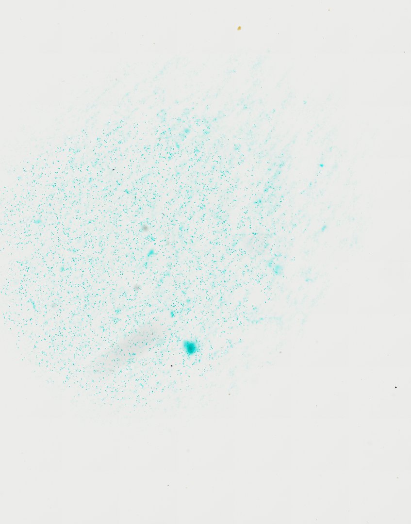
This slide shows many Chlamydomonas cells, a single-celled green alga, stained with fast green. It is a classic example of a simple, swimming photosynthetic cell.
A whole mount (W.M.) preparation of Chlamydomonas (labeled Chlamydomonas W.M.). The slide demonstrates unicellular green algae featuring distinct ovoid cells, cup-shaped chloroplasts, and flagella utilized for motility, illustrating classic chlorophyte morphology.
What to look for
- Cell shape: Small oval to round cells scattered across the slide.
- Cup-shaped chloroplast: One large chloroplast that fills much of the cell, shaped like a cup.
- Pyrenoid: A small round body inside the chloroplast where starch is made.
- Flagella: Two thin whip-like threads at one end, often hard to see on stained slides.
- Eyespot: A small spot near the front of the cell that senses light, not always visible after staining.
At each magnification
Low power (about 4x)
Tiny scattered dots across the slide.
Medium power (about 10x)
Oval cells with a visible outline.
High power (about 40x)
The chloroplast, pyrenoid, and possibly the flagella.
Questions and answers
What does Chlamydomonas look like under a microscope?
Chlamydomonas is a small oval cell, often about 10 micrometers long, with one large cup-shaped chloroplast. It has two flagella at one end for swimming, which can be hard to see without high magnification.
Is Chlamydomonas a plant or an animal?
Neither. Chlamydomonas is a single-celled green alga. It makes its own food by photosynthesis like a plant, but it swims with flagella, which is why it is used to discuss how living things are classified.
- Category
- Botany & Plants
- Scan magnification
- 20x
- Stain
- Fast Green
- Tags
- Chlamydomonas W.M., Chlamydomonas w.m., Chlamydomonas whole mount, Chlamydomonas, Chlamydomonas reinhardtii, Chlorophyta
Viewing this slide in class
This is a scan of a real prepared slide, not a simulation or a drawing. On StudyScope you can pan from the whole slide down to individual structures, always in focus, and measure them in real micrometers with the calibrated ruler. Present it live and every student screen follows your view and annotations. Students join with a room code on Chromebooks, iPads, or any browser, with no accounts.