Rapeseed Flower Bud Cross Section Under the Microscope
Rapeseed Flower Bud (Cross Section), Brassica napus
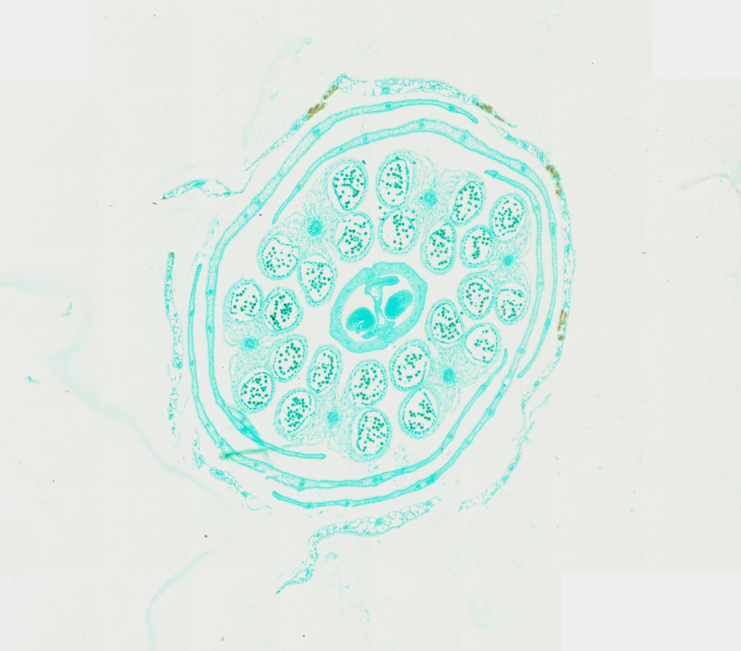
This is a cross section through a rapeseed (canola) flower bud stained with fast green. One slice shows all the floral parts arranged in rings, like a real-life floral diagram.
This transverse section through a rapeseed (Brassica napus) floral bud displays the complete concentric arrangement of floral whorls typical of Brassicaceae. Enclosing the exterior are the outer sepals (calyx) and delicate petals (corolla). Positioned centrally within the whorl are multiple tetradynamous stamens, whose anthers reveal distinct pollen sacs (microsporangia) filled with developing microspores/pollen grains. At the absolute center of the bud section sits the superior ovary, composed of two fused carpels separated by a false septum (replum) with developing ovules attached along parietal placentas. Fast Green counterstaining clearly outlines the cellular architecture, vascular bundles, and protective dermal tissues across all floral organs.
What to look for
- Sepals: The outer ring of protective leaf-like parts.
- Petals: Thin parts just inside the sepals.
- Stamens and anthers: Usually six stamens, with anthers showing chambers full of developing pollen.
- Ovary: The central structure made of two joined carpels.
- False septum: A thin wall dividing the ovary into two chambers.
- Ovules: Small round structures attached along the ovary walls.
At each magnification
Low power (about 4x)
Rings of floral parts around a central ovary.
Medium power (about 10x)
The anthers with their pollen sacs and the two-chambered ovary.
High power (about 40x)
Developing pollen inside the anthers and the ovules in the ovary.
Questions and answers
What does a flower bud cross section look like under a microscope?
A flower bud cross section shows the floral parts in rings: sepals on the outside, then petals, then stamens with anthers, and the ovary in the center. It looks a lot like the floral diagrams in textbooks.
How many parts does a mustard family flower have?
Flowers in the mustard family, like rapeseed, usually have four sepals, four petals, six stamens (four long and two short), and an ovary made of two carpels.
- Category
- Botany & Plants
- Scan magnification
- 20x
- Stain
- Fast Green
- Tags
- rapeseed flower bud cross section, Brassica napus, rapeseed flower, floral bud cross section, anther cross section, pollen sacs
Viewing this slide in class
This is a scan of a real prepared slide, not a simulation or a drawing. On StudyScope you can pan from the whole slide down to individual structures, always in focus, and measure them in real micrometers with the calibrated ruler. Present it live and every student screen follows your view and annotations. Students join with a room code on Chromebooks, iPads, or any browser, with no accounts.