Skeletal Muscle Under the Microscope
Skeletal Muscle (Transverse & Longitudinal Section), Homo sapiens
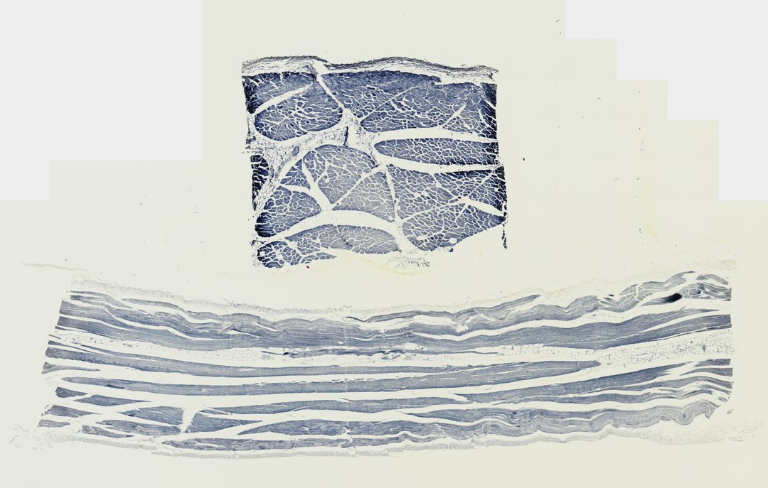
This slide shows skeletal muscle cut two ways, across the fibers and along them, stained with iron hematoxylin. It is the standard slide for seeing muscle fibers, their many nuclei, and the stripes (striations) that make skeletal muscle unique.
A histological preparation showing skeletal muscle in both transverse section (T.S.) and longitudinal section (L.S.). The top section demonstrates cross-sectioned muscle fascicles wrapped in perimysium with individual polygonal muscle fibers bounded by endomysium. The lower section illustrates elongated parallel muscle fibers featuring peripheral multi-nucleation and characteristic cross-striations of myofibrils.
What to look for
- Muscle fibers (cross section): Many-sided shapes packed together, each one a single long muscle cell cut across.
- Fascicles: Bundles of fibers grouped together and wrapped in connective tissue (perimysium).
- Nuclei at the edge: Dark nuclei pressed against the outer edge of each fiber, not in the center.
- Muscle fibers (longitudinal section): Long, parallel fibers lying side by side.
- Striations: Fine alternating light and dark bands across the long fibers, stained dark by iron hematoxylin.
At each magnification
Low power (about 4x)
Two areas of muscle: one of packed polygons (cut across) and one of long stripes (cut lengthwise).
Medium power (about 10x)
Fascicles in the cross section and long parallel fibers in the lengthwise section.
High power (about 40x)
The cross-striations and the many nuclei along the edge of each fiber.
Questions and answers
What does skeletal muscle look like under a microscope?
Skeletal muscle is made of long, cylindrical fibers with many nuclei along their edges. Cut lengthwise, the fibers show fine light and dark stripes called striations. Cut across, they look like packed polygons grouped into bundles.
How do you tell skeletal, cardiac, and smooth muscle apart?
Skeletal muscle has long, striped fibers with many nuclei at the edge. Cardiac muscle is striped too but has branched cells with one central nucleus and dark lines between cells. Smooth muscle has no stripes and is made of spindle-shaped cells with one central nucleus.
Why does skeletal muscle have stripes?
The stripes come from repeating units of the proteins actin and myosin inside each fiber. These units, called sarcomeres, line up across the fiber and slide together when the muscle contracts.
- Category
- Histology and Tissue
- Scan magnification
- 20x
- Stain
- Iron hematoxylin
- Tags
- Skeletal muscle T.S. and L.S., Skeletal muscle cross section, Skeletal muscle longitudinal section, Muscle tissue, Striated muscle, Muscle fascicle
Viewing this slide in class
This is a scan of a real prepared slide, not a simulation or a drawing. On StudyScope you can pan from the whole slide down to individual structures, always in focus, and measure them in real micrometers with the calibrated ruler. Present it live and every student screen follows your view and annotations. Students join with a room code on Chromebooks, iPads, or any browser, with no accounts.
More Histology and Tissue slides
- Bone Cell (Hyaline Cartilage / Trachea Section)
- Lung (bottom section)
- Lung (bottom section)
- Lung (top section)
- Lung (top section)
- Lung (top section)
- Lung (top section)
- Oral Epithelial Cells (Whole Mount)