Small Intestine Cross Section Under the Microscope
Small Intestine (Cross Section)
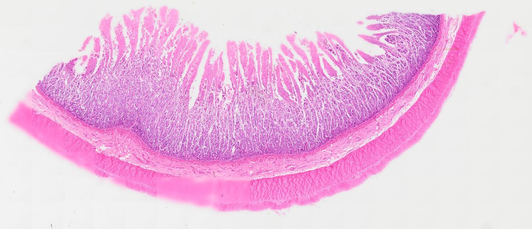
This is a cross section of the small intestine stained with H&E. It shows the finger-like villi that absorb food and the layers that make up the wall of the digestive tract.
This cross-sectional histological preparation displays the characteristic four-layered wall architecture of the small intestine stained with Hematoxylin and Eosin (H&E). Extending into the lumen are tall, finger-like villi lined by simple columnar epithelium consisting primarily of absorptive enterocytes with apical microvilli (striated border) and interspersed mucin-secreting goblet cells. At the base of the villi, tubular invaginations form the crypts of Lieberkühn within the lamina propria. Beneath the mucosa, the thin muscularis mucosae separates the inner layer from the connective tissue submucosa. Surrounding the submucosa is a thick muscularis externa composed of inner circular and outer longitudinal smooth muscle layers, bounded externally by the visceral peritoneum (serosa).
What to look for
- Villi: Finger-like folds sticking into the open center (lumen).
- Simple columnar epithelium: A single layer of tall cells lining each villus, with purple nuclei lined up near their base.
- Goblet cells: Pale, cup-shaped cells among the lining cells that make mucus.
- Crypts: Tube-shaped pockets between the bases of the villi.
- Submucosa: A layer of loose pink connective tissue below the lining.
- Muscularis externa: Two layers of smooth muscle, an inner circular one and an outer longitudinal one.
At each magnification
Low power (about 4x)
A ring-shaped wall with a fringe of villi pointing into the lumen.
Medium power (about 10x)
The villi, the crypts, and the muscle layers of the wall.
High power (about 40x)
Tall lining cells, pale goblet cells, and the brush-like edge of the lining.
Questions and answers
What does the small intestine look like under a microscope?
The small intestine wall has finger-like villi lining the inside, covered by a single layer of tall cells with pale goblet cells mixed in. Below that are the submucosa and two layers of smooth muscle.
Why does the small intestine have villi?
Villi greatly increase the surface area for absorbing nutrients. The cells on the villi also have tiny projections called microvilli, which add even more surface area.
What are the layers of the small intestine wall?
From the inside out, the layers are the mucosa (lining with villi), the submucosa (connective tissue), the muscularis externa (two layers of smooth muscle), and the serosa (a thin outer covering).
- Category
- Histology and Tissue
- Scan magnification
- 20x
- Stain
- H&E
- Tags
- small intestine cross section, small intestine, intestinal villi, crypts of Lieberkühn, simple columnar epithelium, enterocytes
Viewing this slide in class
This is a scan of a real prepared slide, not a simulation or a drawing. On StudyScope you can pan from the whole slide down to individual structures, always in focus, and measure them in real micrometers with the calibrated ruler. Present it live and every student screen follows your view and annotations. Students join with a room code on Chromebooks, iPads, or any browser, with no accounts.
More Histology and Tissue slides
- Bone Cell (Hyaline Cartilage / Trachea Section)
- Lung (bottom section)
- Lung (bottom section)
- Lung (top section)
- Lung (top section)
- Lung (top section)
- Lung (top section)
- Oral Epithelial Cells (Whole Mount)