Sunflower Stem Under the Microscope
Sunflower Stem (Longitudinal Section), Helianthus annuus
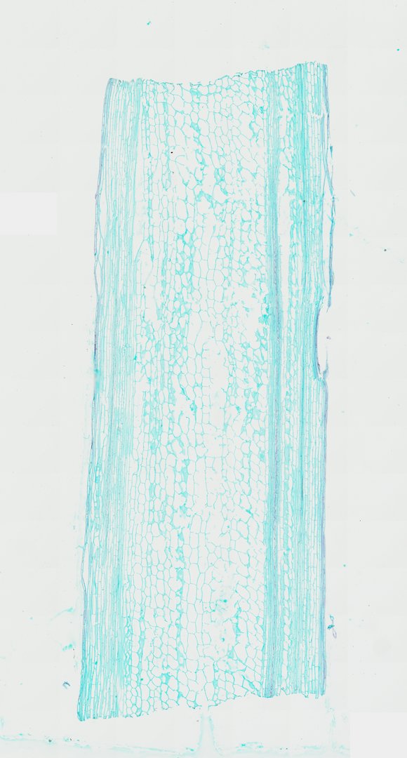
This is a lengthwise section of a sunflower stem stained with safranin and fast green. Cutting along the stem shows xylem vessels as long tubes with spiral and ring patterns in their walls.
This longitudinal section of a sunflower stem illustrates the axial orientation of plant tissues in a typical dicotyledonous stem. Running along the outer flanks are elongated epidermal and cortical collenchyma cells providing structural support. Flanking the central region are prominent vascular strands showing parallel, elongated vascular elements, including lignified xylem vessels with reticulate, annular, and spiral wall thickenings, along with narrow phloem sieve tube members and companion cells. The central axis features an expansive pith (medulla) composed of large, thin-walled parenchyma cells with visible intercellular spaces, characteristic of herbaceous dicot stem anatomy under differential botanical staining.
What to look for
- Epidermis and cortex: Outer layers of long cells running along the edges of the section.
- Xylem vessels: Long tubes stained red, with spiral, ring, or net-like thickenings in their walls.
- Phloem: Narrow, green-stained cells running beside the xylem.
- Pith: Large, thin-walled cells in the center of the stem.
At each magnification
Low power (about 4x)
Long bands of tissue running the length of the section, with red stripes where the xylem is.
Medium power (about 10x)
Long xylem vessels next to narrower phloem cells, and the large pith cells in the middle.
High power (about 40x)
The spiral and ring-shaped wall patterns inside xylem vessels.
Questions and answers
What do xylem vessels look like in a longitudinal section?
In a longitudinal section, xylem vessels look like long tubes. Their walls have red-stained thickenings in patterns such as spirals, rings, or nets, which help keep the tubes from collapsing.
Why do xylem vessels have spiral walls?
Spiral and ring thickenings strengthen the vessel walls while still letting young stems keep growing longer. Later-forming vessels often have more solid walls with pits.
- Category
- Botany & Plants
- Scan magnification
- 20x
- Stain
- Safranin / Fast Green
- Tags
- sunflower stem, Helianthus annuus, longitudinal section, sunflower, dicot stem, vascular bundle
Viewing this slide in class
This is a scan of a real prepared slide, not a simulation or a drawing. On StudyScope you can pan from the whole slide down to individual structures, always in focus, and measure them in real micrometers with the calibrated ruler. Present it live and every student screen follows your view and annotations. Students join with a room code on Chromebooks, iPads, or any browser, with no accounts.