Wheat Seed / Grain Cross Section Under the Microscope
Wheat Seed / Grain (Cross Section), Triticum aestivum
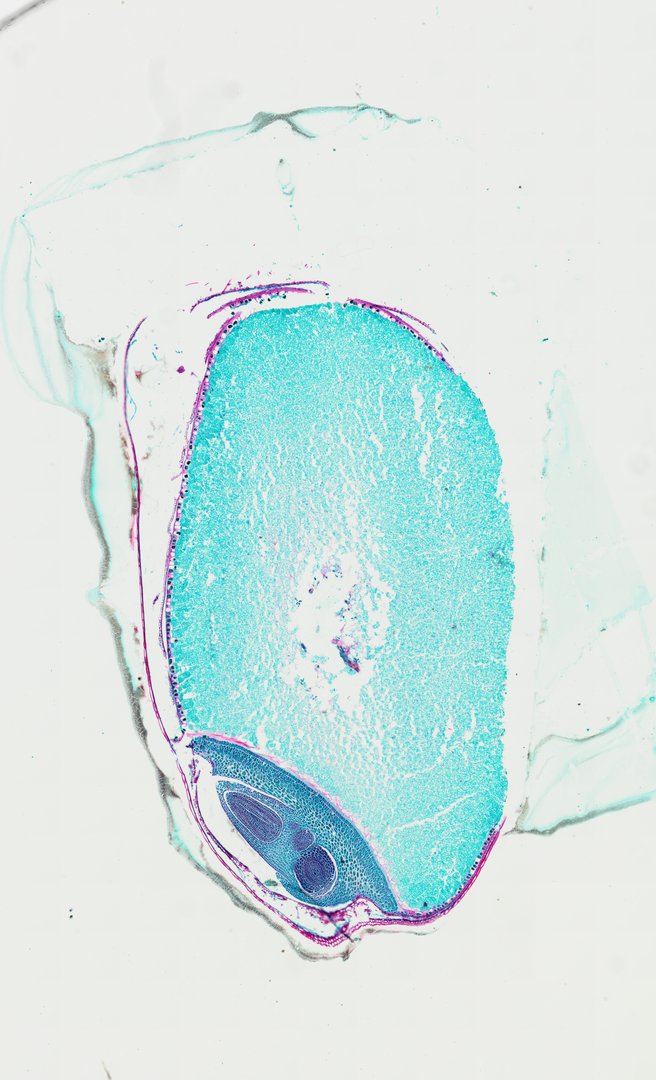
This is a cross section of a wheat grain stained with safranin and fast green. It shows the parts of a cereal grain, which is where flour and bran come from.
A cross section through a wheat grain or caryopsis (Wheat Seed C.S.) double-stained with Safranin and Fast Green. The section reveals the outer protective layer composed of the pericarp fused with the seed coat (testa), bounded internally by a distinct peripheral aleurone layer. The bulk of the interior consists of starchy endosperm cells, while the basal region captures the monocot embryo (germ) including the scutellum and embryonic axis structures. This slide highlights cereal grain anatomy and seed storage tissues characteristic of Poaceae.
What to look for
- Pericarp and seed coat: The thin outer layers of the grain, which make up much of the bran.
- Aleurone layer: A single row of boxy cells just inside the outer layers.
- Starchy endosperm: Large cells filling most of the grain, packed with starch.
- Crease: A groove running along one side of the grain, which shows as a notch in the outline.
- Embryo: The germ, found at the base of the grain, if the section passes through it.
At each magnification
Low power (about 4x)
The rounded grain outline with a notch on one side, mostly filled with endosperm.
Medium power (about 10x)
The outer layers and the row of aleurone cells.
High power (about 40x)
The boxy aleurone cells and the starch-filled endosperm cells.
Questions and answers
What are the parts of a wheat grain?
A wheat grain has outer layers (pericarp and seed coat), an aleurone layer, a large starchy endosperm, and an embryo, or germ. White flour comes mostly from the endosperm, while whole wheat flour also includes the bran and germ.
What does the aleurone layer do?
The aleurone layer is a row of cells around the endosperm. When the seed sprouts, these cells release enzymes that break down the stored starch into sugars the young plant can use.
- Category
- Botany & Plants
- Scan magnification
- 20x
- Stain
- Safranin / Fast Green
- Tags
- wheat seed cross section, Wheat Seed C.S., Triticum aestivum, wheat grain, caryopsis, monocot seed
Viewing this slide in class
This is a scan of a real prepared slide, not a simulation or a drawing. On StudyScope you can pan from the whole slide down to individual structures, always in focus, and measure them in real micrometers with the calibrated ruler. Present it live and every student screen follows your view and annotations. Students join with a room code on Chromebooks, iPads, or any browser, with no accounts.