Cotton Root Cross Section Under the Microscope
Cotton Root (Cross Section), Gossypium hirsutum
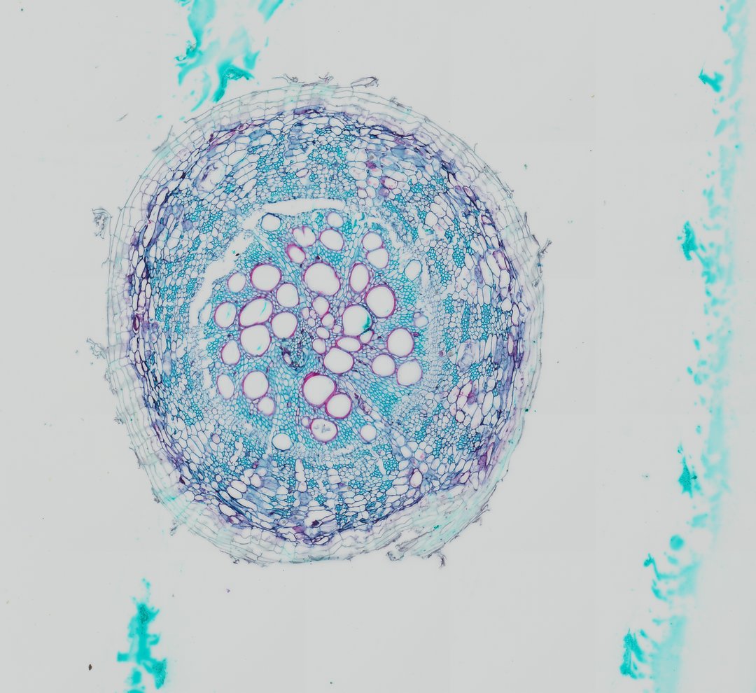
This is a cross section of a young cotton root stained with safranin and fast green. It shows the layers of a dicot root, from the outer skin to the vascular core in the center.
This cross section of a cotton root (Gossypium hirsutum c.s.) reveals the fundamental anatomical organization of a young dicotyledonous root undergoing primary growth and early vascular development. The outermost boundary is composed of an epidermis and underlying cortical parenchyma cells that store reserves and facilitate lateral water transport. Centrally located within the vascular cylinder (stele), a prominent arrangement of xylem vessels displays thick, lignified secondary cell walls stained red/magenta by safranin, interspaced with primary and secondary phloem elements stained blue-green. Surrounding the vascular tissue is the endodermis, which features Casparian strips regulating apoplastic transport into the vascular core. The distinct star-shaped or tetrarch/polyarch vascular arrangement and peripheral pericycle layer highlight structural features characteristic of Malvaceae root anatomy.
What to look for
- Epidermis: The outer layer of small cells around the edge of the root.
- Cortex: A wide zone of large, round, thin-walled cells, stained green, between the epidermis and the center.
- Endodermis: A single ring of cells at the inner edge of the cortex that controls what enters the vascular core.
- Pericycle: A thin layer just inside the endodermis, where side roots begin.
- Xylem: Thick-walled cells stained red in the center, often arranged in a star or cross shape.
- Phloem: Small green-stained cell groups sitting between the arms of the xylem.
At each magnification
Low power (about 4x)
A round section with a wide pale cortex and a small colored core in the center.
Medium power (about 10x)
The endodermis ring and the star-shaped arrangement of red xylem.
High power (about 40x)
Individual xylem vessels with thick red walls and the small phloem cells between them.
Questions and answers
What does a dicot root cross section look like under a microscope?
A dicot root has an epidermis, a wide cortex, a ring of endodermis, and a central core where xylem forms a star or cross shape with phloem between the arms. With safranin and fast green, the xylem is red and most other tissue is green.
What is the endodermis in a root?
The endodermis is a single layer of cells around the vascular core. Its walls have a waterproof band called the Casparian strip, which forces water and minerals to pass through the cells, so the root can control what it takes in.
How is a root cross section different from a stem cross section?
In a dicot root, xylem and phloem alternate around the center, with xylem often in a star shape. In a dicot stem, xylem and phloem sit together in bundles arranged in a ring around a central pith.
- Category
- Botany & Plants
- Scan magnification
- 20x
- Stain
- Safranin and Fast Green
- Tags
- cotton root cross section, Gossypium hirsutum, dicot root, xylem vessels, phloem, stele
Viewing this slide in class
This is a scan of a real prepared slide, not a simulation or a drawing. On StudyScope you can pan from the whole slide down to individual structures, always in focus, and measure them in real micrometers with the calibrated ruler. Present it live and every student screen follows your view and annotations. Students join with a room code on Chromebooks, iPads, or any browser, with no accounts.