Short answer: A leaf cross section shows the leaf as a stack of layers. From top to bottom in a typical dicot leaf: a waxy cuticle, the upper epidermis, tall palisade cells that do most of the photosynthesis, loose spongy cells with air spaces, and the lower epidermis. Veins carrying xylem and phloem run through the middle, and on slides stained with safranin and fast green the thick-walled xylem stains red to purple and most other cells green. Stomata, the small pores flanked by guard cells, are easiest to study face-on in a peel of the lower epidermis, where you can count them and measure them.
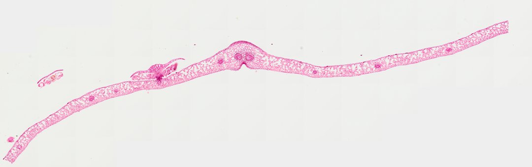
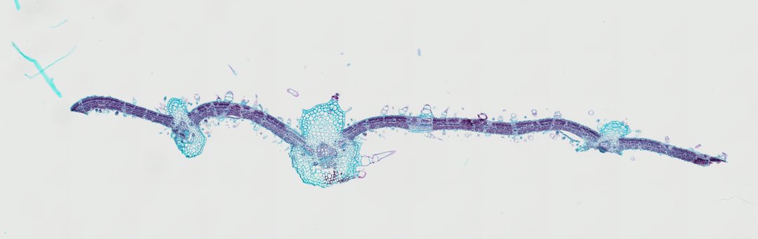 Sunflower Leaf (Cross Section)Helianthus annuusSafranin / Fast Green
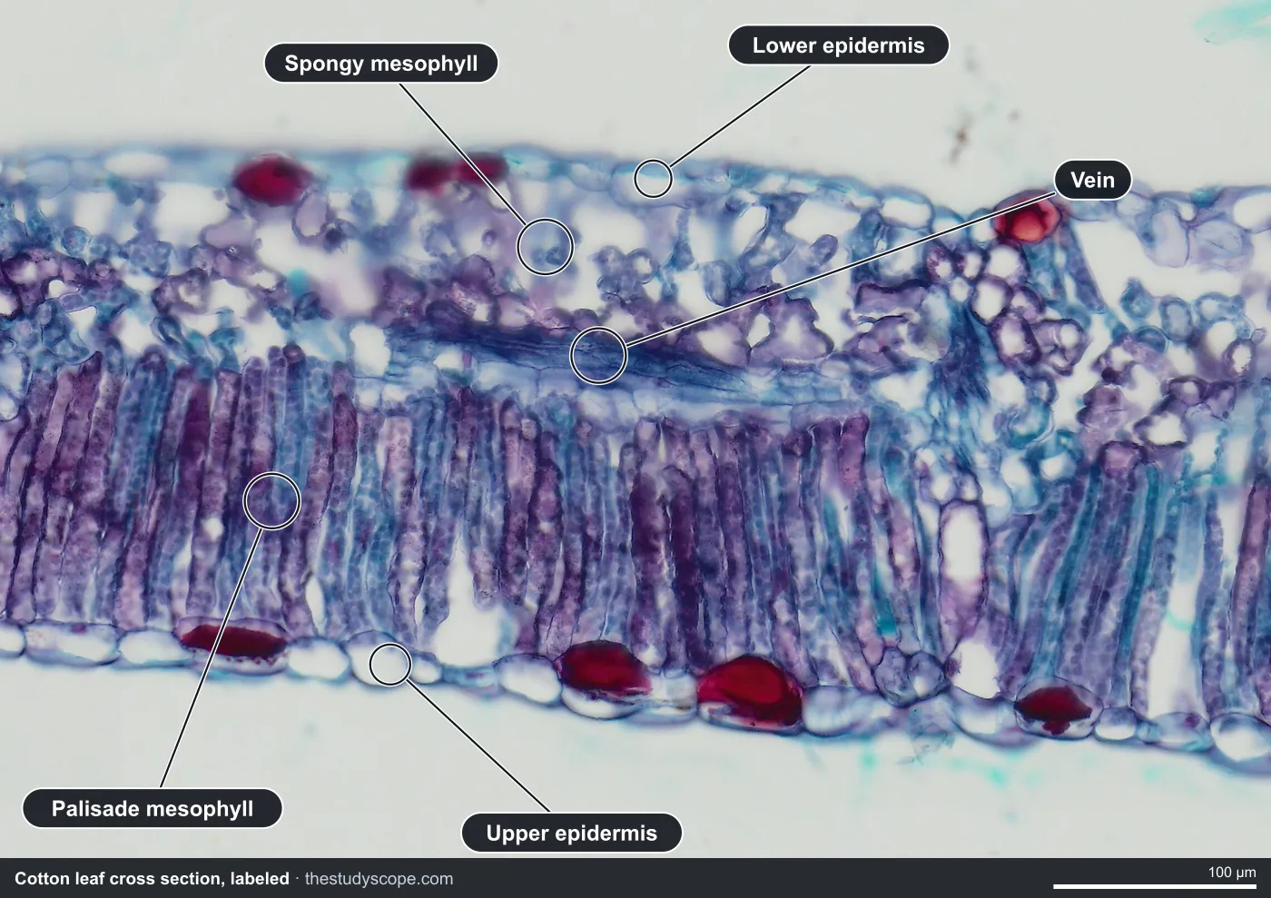
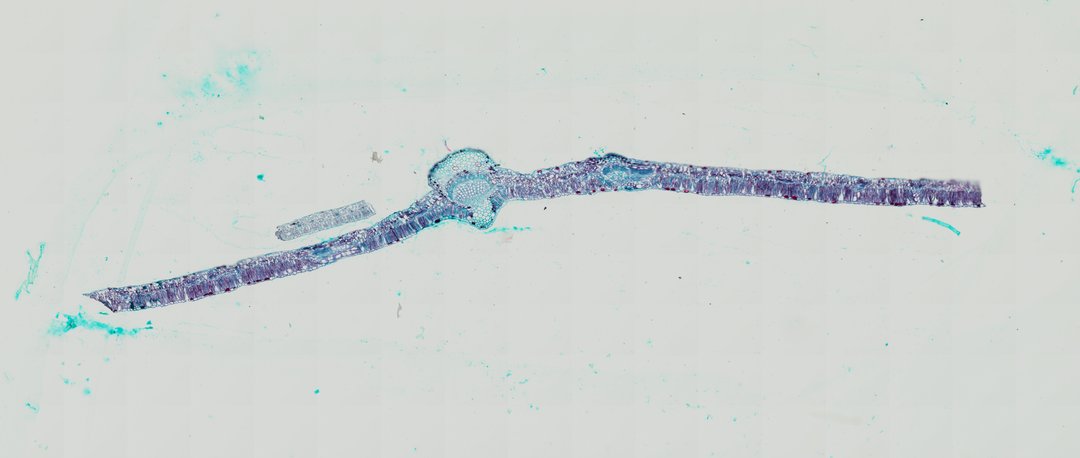 Cotton Leaf (Cross Section)Gossypium hirsutumSafranin / Fast Green
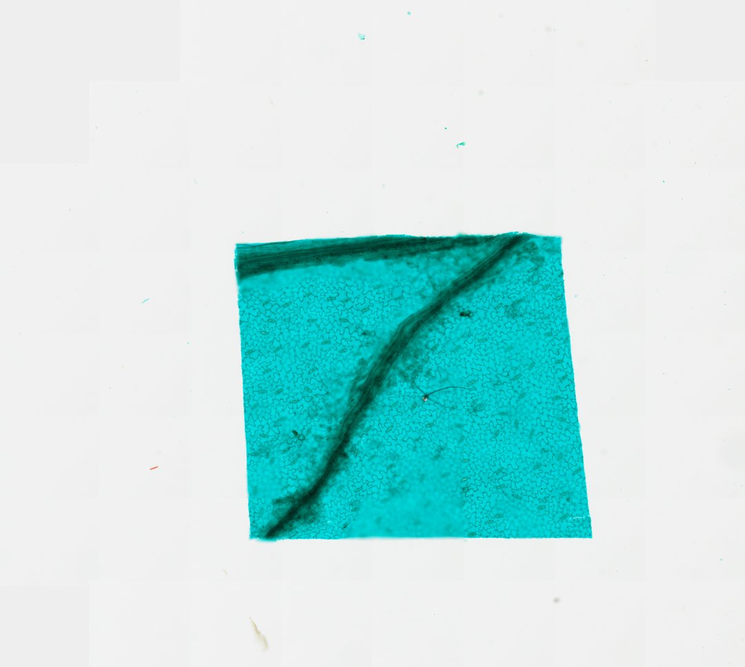 Lower Epidermis of Broad Bean Leaf (Whole Mount)Vicia fabaFast Green
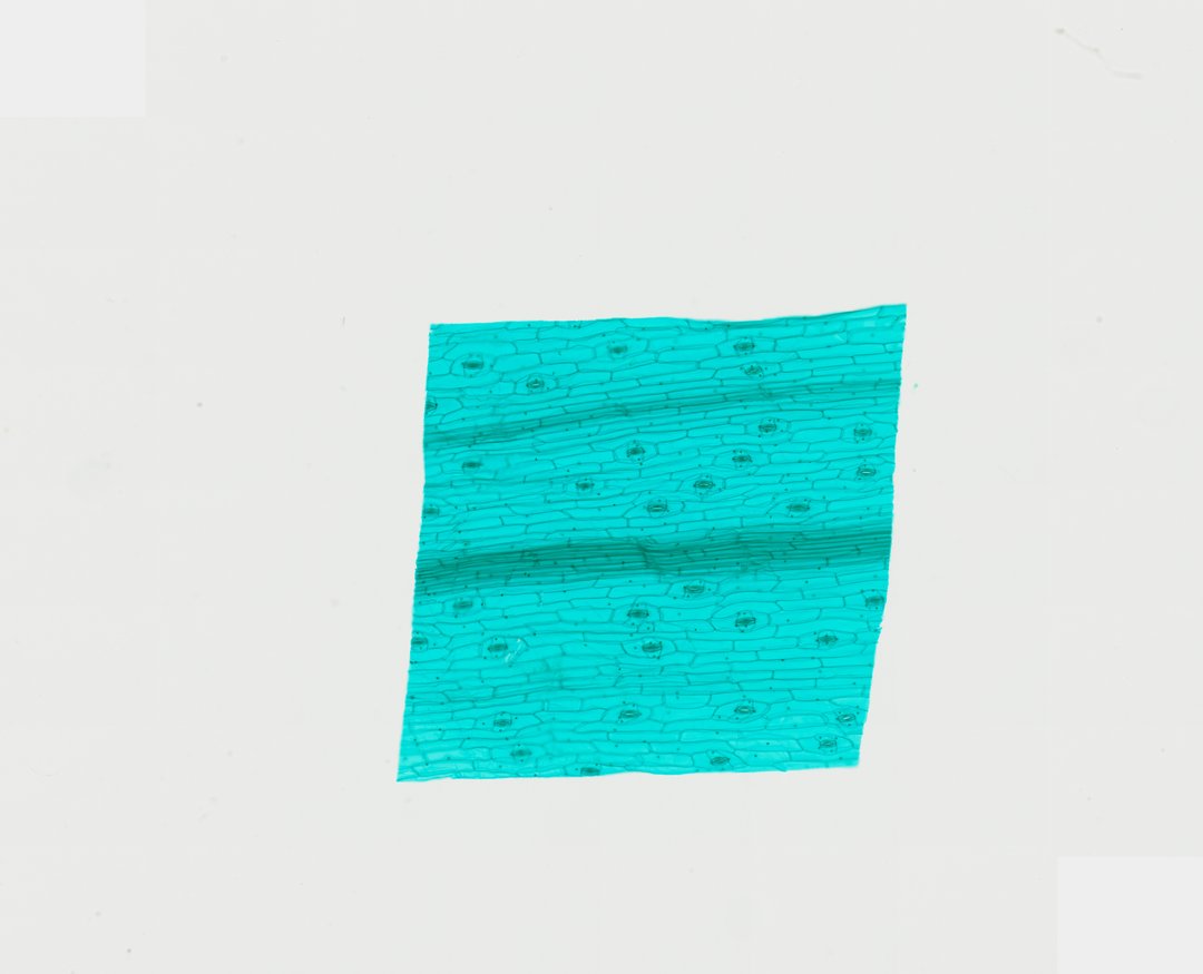 Lower Epidermis of Tradescantia reflexa Leaf (Whole Mount)Tradescantia reflexaFast Green
Searching for a labeled leaf cross section diagram usually turns up drawings. These are real glass slides instead, so you see what a leaf actually looks like, with uneven cells and stain colors. This guide tells you what each layer should look like and where to find it.
The layers of a leaf cross section
| Layer | What it looks like | Its job |
|---|---|---|
| Cuticle | A thin waxy coat on the outside of the epidermis; thicker on leathery or evergreen leaves | Slows water loss |
| Upper epidermis | A single layer of cells across the top of the blade | Protective skin |
| Palisade mesophyll | Tall, tightly packed cells just under the upper epidermis, like fence posts | Catches light; most photosynthesis happens here |
| Spongy mesophyll | Loose, rounded cells with open air spaces, toward the lower surface | Lets carbon dioxide, oxygen, and water vapor move through the leaf |
| Vein (vascular bundle) | Xylem on the upper side and phloem below it (xylem stains red to purple with safranin); the midrib is the biggest vein | Xylem carries water in; phloem carries sugar out |
| Lower epidermis | A single layer of cells along the bottom of the blade, with stomata | Protective skin; gas exchange through the stomata |
| Stomata and guard cells | Small pores, each between two bean-shaped guard cells | Let gases in and out; guard cells open and close the pore |
What to look for in the sunflower and cotton leaves
Both the sunflower leaf and the cotton leaf are dicots stained with safranin and fast green, so the thick-walled xylem stains red to purple and most other cells green. Start at the lowest zoom, where you can see the thin blade and the thick midrib bulging from it, then zoom into the blade and find the palisade layer, which always marks the upper surface. The cotton section lies upside down on its slide, so its palisade layer runs along the bottom of the view.
- Epidermis: a single layer of cells on the top and the bottom of the blade.
- Palisade mesophyll: tall cells packed side by side just under the upper epidermis. This is the layer to find first.
- Spongy mesophyll: looser cells with air spaces, below the palisade layer.
- Midrib: the thick central vein, with a bundle of xylem and phloem near its center.
- Trichomes (sunflower only): hairs made of several cells sticking out from both surfaces.
How to identify xylem and phloem under the microscope
- Find a vein first. Look at the midrib, where the bundle is largest.
- Look for the thick-walled cells, often in rows. Safranin stains their walls red to purple.
- Look on the side toward the lower surface for a patch of small, thin-walled green cells. That is the phloem.
- Check the orientation: in a leaf vein, xylem faces the upper surface and phloem faces the lower. Use the palisade layer to tell which surface is up.
Monocot vs dicot leaf cross section
The lily leaf is a monocot. Its veins run in parallel, so a cross section shows a row of similar vascular bundles, and the mesophyll is not clearly split into palisade and spongy layers the way it is in the sunflower and cotton leaves. It also shows the waxy cuticle, stomata in the epidermis, and red xylem above green phloem in each bundle. Put it next to the sunflower leaf and ask students what is different. For the stem version of this comparison, see our guide to monocot vs dicot stem.
Leaves built for dry or cold places
Not every leaf follows the textbook layout. Several slides show how leaves change to save water:
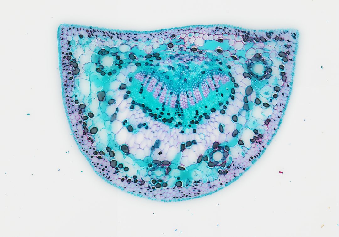
- Pine needle (a good one for pine needle cross section labeling practice): a thick waxy cuticle, a tough layer of thick-walled cells under the skin, sunken stomata, mesophyll cells with folded walls, round resin ducts, and a central vascular region wrapped in an endodermis.
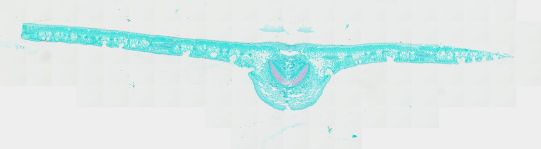
- Oleander (Nerium): a heavy cuticle over several layers of epidermis, tall palisade cells, and hair-lined pits on the underside called stomatal crypts that hold the stomata.
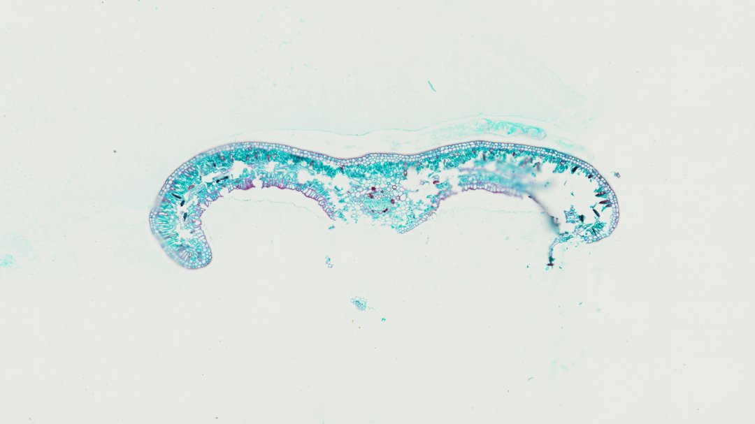
- Sago palm (Cycas): a stiff leaflet with edges that curl down and under, a thick cuticle, a red-stained hypodermis, palisade and spongy layers, and one central vein.
- Fern leaf: a thin blade with a single epidermis on each side and loose mesophyll between them. It also shows a sorus, a cluster of spore cases on the underside, since ferns spread by spores rather than seeds.
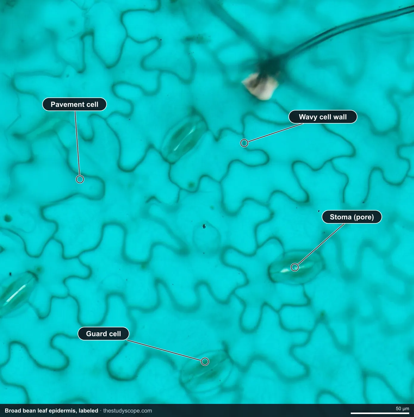
Stomata under the microscope, labeled
A cross section cuts through the leaf, so a stoma shows up only as a gap in the epidermis. To see stomata the way most people picture them, look straight down on the epidermis. These two whole-mount peels do that, so you can find stomata and guard cells at the same zoom level in two different plants.
| Feature | Broad bean | Tradescantia |
|---|---|---|
| Epidermal (pavement) cells | Wavy walls that fit together like puzzle pieces | Long rectangles lined up in parallel rows |
| Guard cells | Pairs of bean-shaped (kidney-shaped) cells around each pore | A pair of cells directly around each pore |
| Helper cells | None; the guard cells sit directly among the pavement cells | Usually four subsidiary cells around each pair of guard cells |
| Other things to spot | Dark strands of a leaf vein crossing the peel | Stomata scattered along the rows |
What to look for in the epidermis peels
- Broad bean, low zoom: a green-stained sheet with a scattering of small dots. Each dot is a stoma.
- Broad bean, medium zoom: the wavy puzzle-piece cells and pairs of guard cells.
- Broad bean, high zoom: the pore between the two kidney-shaped guard cells.
- Tradescantia, medium zoom: the rectangular cells and the ring of subsidiary cells around each stoma.
- Tradescantia, high zoom: the guard cells and pore at the center of each stomatal complex.
A prepared slide does not replace the classic hands-on stomata lab. Painting clear nail polish on a leaf, letting it dry, and lifting the film with clear tape gives students a peel of their own plant to look at on a bench microscope. Use the slides here alongside it: they are sharper and stained, and every student can look at the same peel at once.
Teaching it: a stomata lab with real measurements
- Open the broad bean peel at low zoom and have students count the stomata in one field of view. Every slide is calibrated, so you can compare the same-size area across slides.
- Zoom in on one stoma and use the ruler to measure a guard cell in micrometers. The ruler reads real micrometers at any zoom.
- Open the Tradescantia peel and ask students to compare the cell shapes and the cells around the guard cells.
- Have students draw a box around one stoma and save a stamped snapshot of their view for a lab notebook or portfolio.
- Finish in a cross section, such as the cotton leaf, and ask where in the layers the stomata would sit and why the spongy air spaces are right behind them.
You can present these slides live: students join with a room code, with no student accounts, and follow on their own screens while your annotations appear. Leaves also appear in the premade lesson Comparative Plant Anatomy: Monocot vs. Dicot, which covers stems, roots, and leaves.
Start with the sunflower leaf for the layers, then open the broad bean peel for stomata. You can browse all of them in the slide catalog without an account.
Questions and answers
What are the layers of a leaf cross section?
From top to bottom, a dicot leaf has a waxy cuticle, an upper epidermis, a palisade mesophyll of tall packed cells, a spongy mesophyll with air spaces, and a lower epidermis. Veins with xylem and phloem run through the middle.
How do you tell xylem from phloem under a microscope?
On slides stained with safranin and fast green, the thick xylem walls stain red to purple and the thin-walled phloem stains green. In a leaf vein, the xylem is on the side facing the upper surface and the phloem is below it.
What do stomata and guard cells look like under a microscope?
In a leaf peel seen from above, a stoma is a small pore between two guard cells. In bean leaves the guard cells are bean-shaped, and the pores are scattered among puzzle-piece shaped epidermal cells.
Why are there more stomata on the bottom of a leaf?
The underside is shaded and cooler, so less water is lost through stomata there. Many land plants have most of their stomata on the lower surface for this reason.
How is a monocot leaf different from a dicot leaf in cross section?
A dicot leaf usually has a clear palisade layer on top and a spongy layer below, with a net of veins. A monocot leaf like lily has parallel veins, so a cross section shows a row of similar bundles, and the mesophyll is less clearly divided.
How are pine needles adapted to save water?
Pine needles have a small surface area, a thick waxy cuticle, a tough layer of thick-walled cells under the skin, and stomata sunk below the surface. Together these reduce water loss in dry or freezing weather.
More guides
- Monocot vs dicot stems compared
- Frog, human, chicken and fish blood compared
- Virtual microscope, simulator or digital microscope?
- Using a microscope on a Chromebook
- How to identify the stages of mitosis in an onion root tip
- Cheek cells vs onion cells
- Pond water organisms identification guide
- How to identify tissue types
- How to measure cell size
- Lily, pine and luffa pollen compared
- Insect mouthparts compared
- Worm body cavities compared