Short answer: A dicot root cross section has three main zones. From the outside in: a thin epidermis, a wide cortex of large round cells, and a small vascular core (the stele) in the center, wrapped in a ring of cells called the endodermis. Inside the core, xylem and phloem sit in separate groups that take turns around the center, instead of sharing bundles the way they do in a stem. As a dicot root gets older, xylem fills the core and the root can grow thicker, which is how a radish gets its size.
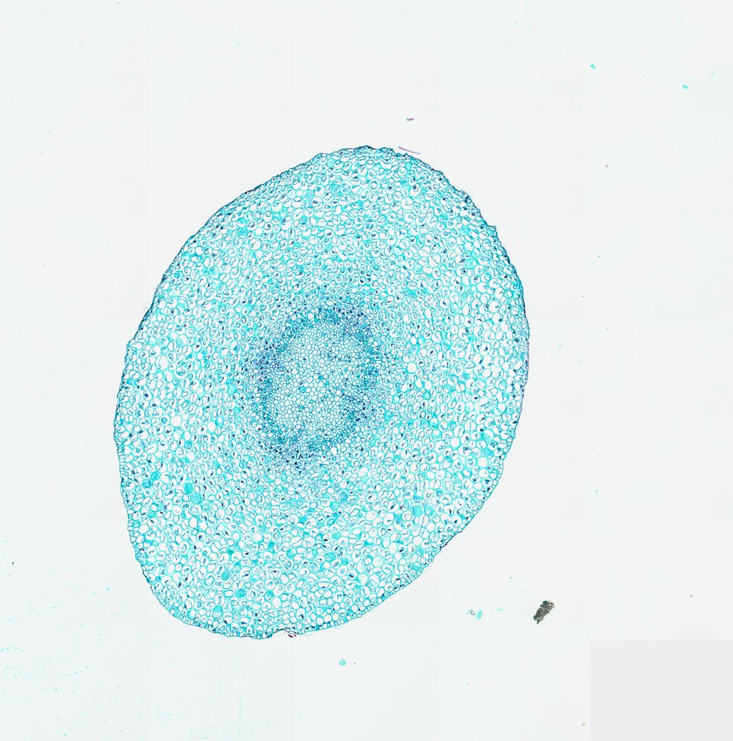 Young Root of Broad Bean (Cross Section)Vicia fabaFast Green
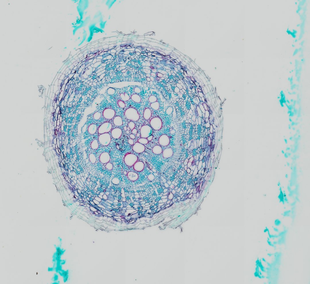 Cotton Root (Cross Section)Gossypium hirsutumSafranin and Fast Green
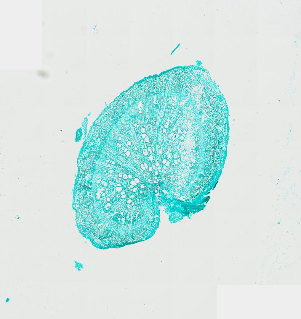 Radish Root (Cross Section)Raphanus sativusFast Green
Labeled dicot root cross section
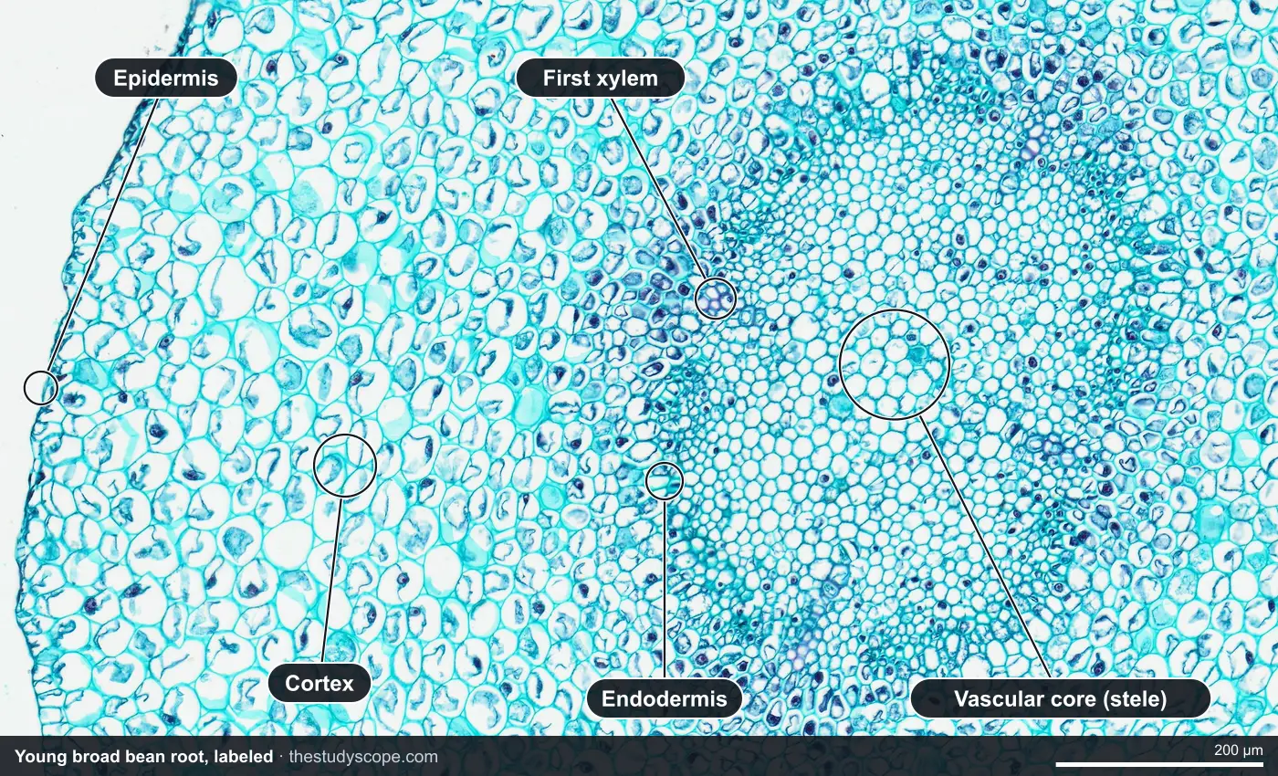
The young broad bean root is the one to learn the layers on. It is about 2 millimeters across at its widest, and most of that is cortex. The core in the middle is only about half a millimeter wide. Many textbooks use a buttercup (Ranunculus) root for this diagram; the broad bean shows the same plan.
Parts of a root cross section, from the outside in
- Epidermis: one thin layer of cells at the edge. In living roots, some of these cells grow root hairs that take in water. On the bean slide it is thin and a little torn, which is normal for a cut root.
- Cortex: the widest zone. Large, round, thin-walled cells with small gaps between them. They store starch and pass water inward toward the core.
- Endodermis: a single ring of cells at the inner edge of the cortex. It works as a checkpoint for everything entering the core (more on that below).
- Pericycle: a thin layer just inside the endodermis. Side roots start here and push out through the cortex.
- Xylem: carries water and minerals up. In a young root the first xylem forms in small groups at the outer edge of the core, and later xylem fills in toward the center.
- Phloem: carries sugar down from the leaves. Its groups sit between the xylem groups, never in front of them.
Three roots, three ages
Looking at one root teaches the layers. Looking at three in a row shows how a dicot root changes as it grows, which is the part students usually miss.
| Feature | Young broad bean root | Cotton root | Radish root |
|---|---|---|---|
| Section size | About 1.7 by 2.1 mm | About 1.7 mm | About 2.2 by 2.6 mm |
| Stain | Fast green | Safranin and fast green | Fast green |
| What fills most of it | Cortex | Xylem | Xylem mixed with storage cells |
| The core | Small, about 0.5 mm, mostly thin-walled cells with a few small xylem groups at its edge | A wide core of large xylem vessels with red walls, some over 0.1 mm across | Rows of xylem vessels fanning out from the center like spokes |
| Growth stage | Primary growth only | Older, with extra xylem added | Secondary growth: a cambium keeps adding cells and the root swells |
Young broad bean root: the layers
At low power the bean root is a pale blue-green disk with a darker spot in the middle. Zoom into that spot and the cells shrink sharply where the cortex ends and the core begins. At the edge of the core are a few small clusters of thick-walled cells with a purple tint: the first xylem. Most of the core is still thin-walled cells, because in a root the xylem forms from the outside in. Botanists call that exarch xylem. Ask students to count the xylem groups around the core before you tell them what they are.
Cotton root: the core takes over
The cotton root is double stained, and the difference is striking. Lignified walls take the red safranin, so the xylem vessels show up as bright red rings, many of them wide open tubes. Green-stained cells pack the spaces between them. The core is no longer a small spot; it is most of the root. The outer layers are loose and partly torn away. Put it next to the bean root and ask: where did all that xylem come from?
Radish root: a root built for storage
The radish root is a young radish, not the full-size one from the store, but it already shows how a taproot swells. Xylem vessels run in rows from the center out toward the edge, and between them are many thin-walled storage cells. Near the outside is a band of small cells lined up in neat rows. That is the cambium zone, where new cells keep forming. Every layer it adds makes the root wider, and the storage cells are the part you eat.
Monocot vs dicot root
| Feature | Dicot root (bean, cotton, radish) | Monocot root (corn, onion, grasses) |
|---|---|---|
| Xylem groups | Few, often 2 to 6, sometimes meeting in a star or cross shape | Many, often more than 6, in a ring |
| Center of the root | Little or no pith; xylem fills the middle as the root ages | A clear pith of thin-walled cells in the middle |
| Growing wider | Can add a cambium and grow thick (radish, carrot, tree roots) | No cambium, so roots stay about the same width |
| Root system | Often one main taproot with side roots | Many thin roots of similar size (fibrous) |
A note on our catalog: all three cross sections here are dicots. Our monocot root slide is a corn root tip cut lengthwise, which shows the root cap and the zones of growth rather than the ring of xylem. For the tip itself, see onion root tip mitosis.
Root vs stem cross section
| Feature | Dicot root | Dicot stem |
|---|---|---|
| Where the vascular tissue is | One core in the center | Bundles in a ring, with pith inside |
| Xylem and phloem | In separate groups that alternate around the center | Together in each bundle, phloem on the outside |
| Oldest xylem | On the outside of the core (exarch) | On the inside of each bundle (endarch) |
| Endodermis | A clear ring around the core | Usually hard to see or missing |
| Surface | No waxy cuticle; root hairs near the tip | Cuticle, and sometimes stomata |
Why the difference? A stem has to resist bending in the wind, and pipes and fibers near the edge make it stiff, like a hollow tube. A root has to resist being pulled, and a strong cord down the middle is better for that. To see the stem side of the comparison, open the sunflower stem or read monocot vs dicot stems.
The endodermis and the Casparian strip
Water can soak into a root two ways: through the cells, or around them through the walls and the gaps between cells. The endodermis closes the second route. Each of its cells has a band of waxy suberin, the Casparian strip, running around its side walls like a gasket. Water and minerals that reach the endodermis have to go through a living cell, which lets the plant choose what gets into the xylem. The strip is too thin to pick out on these slides, so look for the ring of cells it belongs to: on the bean root, that ring sits where the big cortex cells suddenly give way to the small cells of the core.
Using these slides in class
- Open all three roots at the lowest zoom and have students sketch where the darker core is in each one, and how much of the root it takes up.
- Zoom into the bean root's core and count the xylem groups. Then compare with the star or cross of xylem students may have seen in a textbook diagram.
- Use the ruler to measure the core and the whole root on the bean and the cotton slides. Every slide is calibrated, so the numbers are real micrometers at any zoom. Work out what fraction of each root is core.
- On the radish, follow one row of vessels from the center to the edge, and find the band of small cells where the root is still growing.
- Finish with a stem students have already seen and ask them to name three ways it differs from the roots.
You can present the slides live: students join with a room code, with no student accounts, and follow your view on their own screens while your annotations appear on theirs.
Want it ready to teach? The premade lab Comparative Plant Anatomy: Monocot vs. Dicot uses all three of these roots plus the corn root tip, along with stems and leaves, and asks students to classify each slide from the evidence. It runs about 50 to 60 minutes. Deep zoom on the slides needs a teacher seat, and you can browse the catalog without an account.
Questions and answers
What are the parts of a root cross section?
From the outside in: the epidermis, a wide cortex, the endodermis (a single ring of cells), the pericycle just inside it, and the vascular core with xylem and phloem. In a young dicot root the xylem and phloem form separate groups that alternate around the center.
What does a dicot root look like under a microscope?
At low power it is a round section with a wide, pale cortex and a small, darker core in the center. Zoomed in, the core has a few groups of thick-walled xylem cells, often arranged like a star or cross, with phloem between them. Older dicot roots have much more xylem and can be mostly core.
What is the difference between a monocot and a dicot root?
A dicot root has a few xylem groups and little or no pith, and many dicot roots can grow thicker with a cambium. A monocot root has many xylem groups in a ring around a clear pith, and no cambium, so it stays thin.
Why is the xylem in the center of a root?
A root is pulled on more than it is bent, and a strong core down the middle resists pulling well. Keeping the vascular tissue in the center also puts it behind the endodermis, which controls what water and minerals reach the xylem.
What is the Casparian strip?
A band of waxy suberin in the side walls of each endodermis cell. It blocks water from slipping between the cells, so water and minerals have to pass through living cells before they reach the xylem. That lets the root filter what it takes in.
Is the xylem in a root exarch or endarch?
Exarch. In a root the first xylem forms at the outer edge of the core and later xylem forms toward the center. Stems are the opposite (endarch), with the first xylem on the inner side of each bundle.
More guides
- Monocot vs dicot stems compared
- Frog, human, chicken and fish blood compared
- Virtual microscope, simulator or digital microscope?
- Using a microscope on a Chromebook
- How to identify the stages of mitosis in an onion root tip
- Cheek cells vs onion cells
- Leaf layers and stomata guide
- Pond water organisms identification guide
- How to identify tissue types
- How to measure cell size
- Lily, pine and luffa pollen compared
- Insect mouthparts compared
- Worm body cavities compared
- Lung alveoli and gills compared
- Insect wings compared