Fern Leaf Cross Section Under the Microscope
Fern Leaf (Cross Section), Polypodiopsida
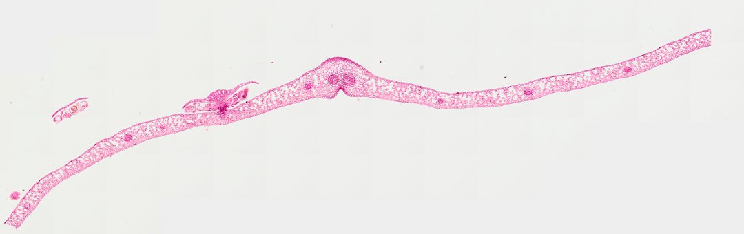
This is a cross section of a fern leaf (frond) stained with safranin. It shows the thin leaf blade and, on the underside, the spore-making structures that ferns use instead of seeds.
A cross section of a fern frond or pinna (Fern leaf Sec.) stained red/pink, illustrating typical pteridophyte leaf anatomy and reproductive structures. The thin leaf blade (lamina) features single-layered upper and lower epidermal cell layers enclosing a mesophyll composed of photosynthetic parenchyma cells with abundant intercellular air spaces. Small vascular bundles with thick-walled xylem elements and phloem tissue are distributed along the blade length, with a central midrib vein providing structural support. On the abaxial surface, a sorus is visible, protected by an indusium flap and containing multiple sporangia filled with developing spores, characteristic of leptosporangiate fern reproduction.
What to look for
- Epidermis: Single layers of cells on the upper and lower surfaces of the leaf.
- Mesophyll: Photosynthetic cells between the two surfaces, with air spaces between them.
- Vascular bundle: A vein with thick-walled xylem cells, stained deep red, and phloem around it.
- Sorus: A cluster of spore cases on the underside of the leaf.
- Sporangia: Small rounded spore cases, each with a row of thickened cells (the annulus) that helps fling spores out.
- Indusium: A thin flap that covers and protects the sorus in many ferns.
At each magnification
Low power (about 4x)
The thin leaf blade with thicker areas where veins and sori are.
Medium power (about 10x)
Clusters of sporangia hanging from the underside of the leaf.
High power (about 40x)
Spores inside the sporangia and the thick-walled cells of the annulus.
Questions and answers
What are the brown dots on the underside of a fern leaf?
They are sori, clusters of sporangia (spore cases). Ferns reproduce with spores rather than seeds, and each sporangium releases many tiny spores.
What does a fern sorus look like under a microscope?
In a cross section, a sorus looks like a cluster of small round capsules on stalks attached to the underside of the leaf. Each capsule is a sporangium full of spores, often covered by a thin flap called the indusium.
- Category
- Botany & Plants
- Scan magnification
- 20x
- Stain
- Safranin
- Tags
- Fern leaf cross section, fern leaf, fern frond, pteridophyte leaf, leaf cross section, sorus
Viewing this slide in class
This is a scan of a real prepared slide, not a simulation or a drawing. On StudyScope you can pan from the whole slide down to individual structures, always in focus, and measure them in real micrometers with the calibrated ruler. Present it live and every student screen follows your view and annotations. Students join with a room code on Chromebooks, iPads, or any browser, with no accounts.