Lily Leaf Cross Section Under the Microscope
Lily Leaf (Cross Section), Lilium
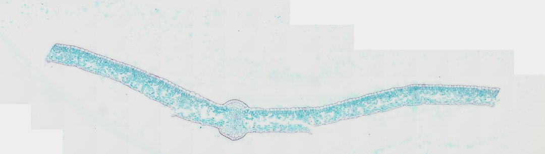
This is a cross section of a lily leaf stained with safranin and fast green. It is a good example of a monocot leaf, where veins run in parallel and the inner cells are not clearly split into palisade and spongy layers.
This cross section of a lily leaf (Lilium leaf c.s.) illustrates classic monocotyledonous foliar architecture under light microscopy. The leaf is bounded by single-layered upper and lower epidermises coated with a protective waxy cuticle, with stomatal complexes distributed across the surfaces to facilitate gas exchange. Internal to the epidermis lies the mesophyll tissue, which in Lilium displays an isobilateral or weakly differentiated arrangement dominated by spongy parenchyma interspersed with intercellular air spaces for efficient gas diffusion during photosynthesis and transpiration. At the center of the blade, a prominent midrib vascular bundle contains oriented xylem vessels positioned adaxially for water transport and phloem elements located abaxially for photosynthate translocation. The surrounding bundle sheath parenchyma provides structural support and mediates transport between the mesophyll cells and vascular tissues.
What to look for
- Epidermis and cuticle: Single layers of cells on both surfaces, coated with a waxy cuticle.
- Stomata: Small pores with guard cells in the epidermis.
- Mesophyll: Photosynthetic cells filling the leaf, with air spaces, and little difference between upper and lower layers.
- Vascular bundles: Veins in a row across the leaf, from its parallel vein pattern.
- Xylem and phloem: Xylem stained red on the upper side of each bundle, phloem stained green below it.
At each magnification
Low power (about 4x)
The leaf blade with a row of vascular bundles across it.
Medium power (about 10x)
Individual bundles with red xylem and green phloem.
High power (about 40x)
Guard cells in the epidermis and the cells around each bundle.
Questions and answers
What is the difference between a monocot and dicot leaf under a microscope?
Dicot leaves usually have a clear palisade layer on top and a spongy layer below, with a net of veins. Monocot leaves like lily have parallel veins, so a cross section shows a row of similar bundles, and the mesophyll is often less clearly divided.
Why is xylem red on this lily leaf slide?
Safranin stains the lignin in xylem walls red, while fast green stains other cell walls green. That makes the water-carrying xylem easy to spot in each vein.
- Category
- Botany & Plants
- Scan magnification
- 20x
- Stain
- Safranin and Fast Green
- Tags
- lily leaf cross section, Lilium, monocot leaf, Lilium leaf c.s., mesophyll, spongy parenchyma
Viewing this slide in class
This is a scan of a real prepared slide, not a simulation or a drawing. On StudyScope you can pan from the whole slide down to individual structures, always in focus, and measure them in real micrometers with the calibrated ruler. Present it live and every student screen follows your view and annotations. Students join with a room code on Chromebooks, iPads, or any browser, with no accounts.