Lower Epidermis of Broad Bean Leaf Under the Microscope
Lower Epidermis of Broad Bean Leaf (Whole Mount), Vicia faba
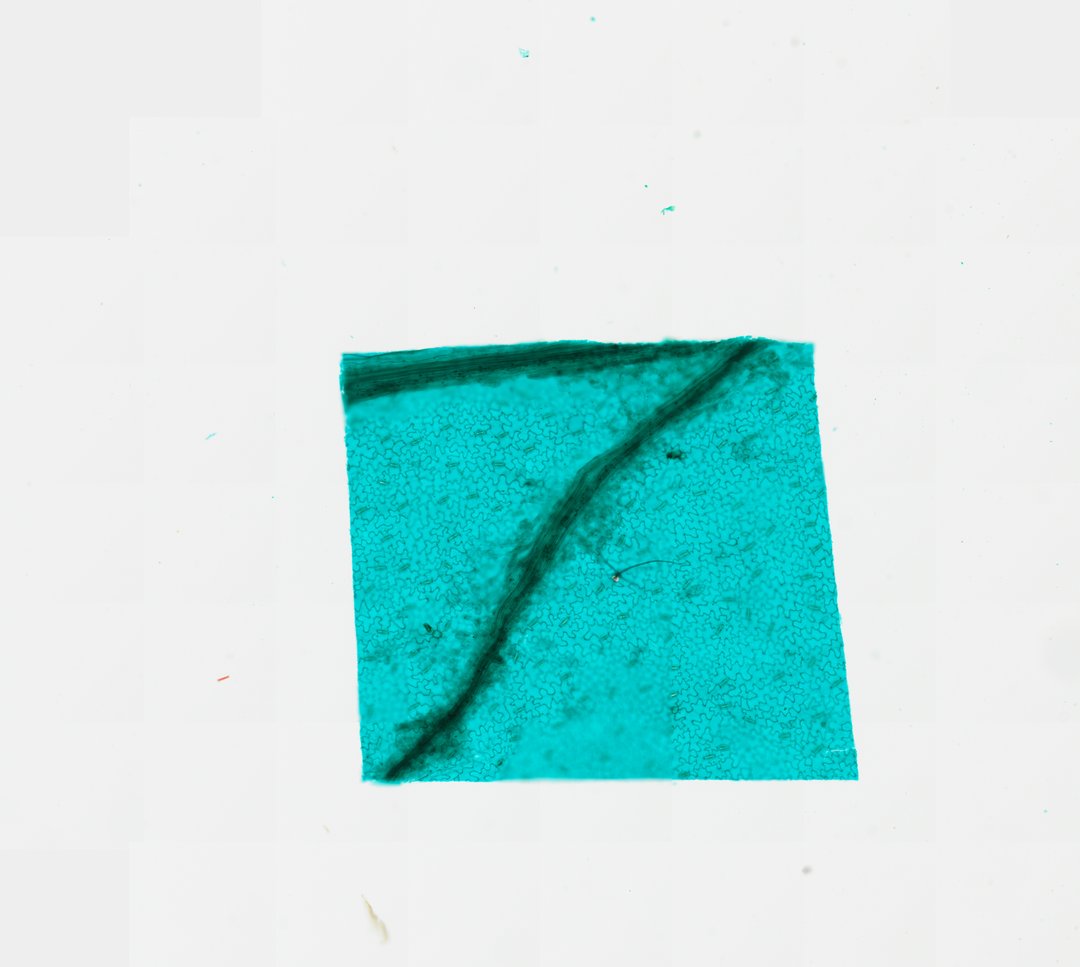
This is a peel of the lower surface of a broad bean leaf, mounted flat and stained with fast green. It is one of the best slides for seeing stomata and the guard cells that open and close them.
This whole mount preparation of the lower epidermis (abaxial surface) of a broad bean (Vicia faba) leaf provides a clear view of dermal tissue adaptation for gas exchange and transpirational regulation. The epidermal layer consists of irregularly shaped, puzzle-piece-like pavement cells with sinuous cell walls interlocked in a continuous monolayer. Embedded across this epidermal sheet are numerous stomatal complexes, each composed of a central stomatal pore bounded by a pair of kidney-shaped (reniform) guard cells. Vascular trace strands run through the field, while Fast Green staining vividly highlights the primary cellulosic walls and structural contours of the epidermal cells and stomatal apparatus.
What to look for
- Pavement cells: Epidermal cells with wavy walls that fit together like puzzle pieces.
- Stomata: Small pores scattered across the surface.
- Guard cells: Pairs of kidney-shaped cells around each pore.
- Veins: Long, narrow cells marking where veins run under the epidermis.
At each magnification
Low power (about 4x)
A green-stained sheet with a scattering of small dots, which are stomata.
Medium power (about 10x)
The wavy puzzle-piece cells and pairs of guard cells.
High power (about 40x)
The pore between the guard cells and the thickened walls around it.
Questions and answers
What do stomata look like under a microscope?
A stoma looks like a small slit or pore between two bean-shaped guard cells. On a leaf peel, they are scattered among the puzzle-piece shaped epidermal cells.
Why are there more stomata on the bottom of a leaf?
The underside of a leaf is shaded and cooler, so less water is lost through stomata there. Many land plants have most of their stomata on the lower surface for this reason.
How do guard cells open and close a stoma?
When guard cells take in water, they swell and bow apart, opening the pore. When they lose water, they go limp and the pore closes, which helps the plant save water.
- Category
- Botany & Plants
- Scan magnification
- 20x
- Stain
- Fast Green
- Tags
- lower epidermis of broad bean leaf, Vicia faba, broad bean epidermis, leaf epidermis whole mount, stomata, guard cells
Viewing this slide in class
This is a scan of a real prepared slide, not a simulation or a drawing. On StudyScope you can pan from the whole slide down to individual structures, always in focus, and measure them in real micrometers with the calibrated ruler. Present it live and every student screen follows your view and annotations. Students join with a room code on Chromebooks, iPads, or any browser, with no accounts.