Mature Anther of Lilium Under the Microscope
Mature Anther of Lilium (Transverse Section), Lilium
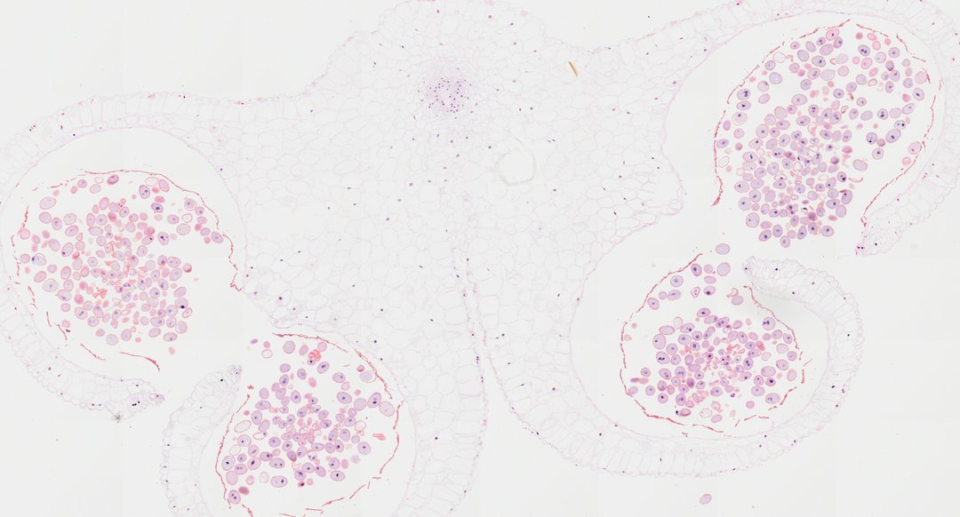
This is a cross section of a mature lily anther stained with safranin and fast green. It shows the four pollen sacs where pollen develops, and it is the standard slide for learning how flowers make pollen.
A transverse section of a mature lily anther (Lilium T.S.) stained with safranin and fast green, displaying the characteristic tetrasporangiate structure typical of angiosperm stamen development. Four prominent pollen sacs (microsporangia) are paired within two anther thecae connected by a central connective tissue containing a single vascular bundle. The anther wall consists of an outer epidermis and a underlying endothecium layer featuring prominent fibrous wall thickenings that aid in dehiscence. Within the spacious pollen chambers, mature, binucleate pollen grains (microspores) with sculptured exine walls are densely packed, having fully developed following meiosis and the degradation of the nourishing tapetal layer.
What to look for
- Pollen sacs: Four chambers arranged in two pairs, making a butterfly-like outline.
- Pollen grains: Many round grains packed inside the sacs.
- Connective tissue: The central tissue joining the two halves of the anther, with a small vascular bundle.
- Epidermis: The outer layer of cells around the anther.
- Endothecium: A layer under the epidermis with thickened walls that help the anther split open.
At each magnification
Low power (about 4x)
The four-lobed shape of the anther with pollen-filled chambers.
Medium power (about 10x)
Pollen grains inside the sacs and the layers of the anther wall.
High power (about 40x)
The textured outer wall of individual pollen grains and the thickened cells of the endothecium.
Questions and answers
What does a lily anther cross section look like under a microscope?
A lily anther cross section shows four pollen sacs arranged in two pairs, joined in the middle by connective tissue with a vein. In a mature anther, the sacs are packed with pollen grains.
Why is lily used for anther slides?
Lily flowers are large, and their anthers and pollen grains are big and easy to section and stain. That makes the stages of pollen development easy to see.
- Category
- Botany & Plants
- Scan magnification
- 20x
- Stain
- Safranin / Fast Green
- Tags
- mature anther of Lilium, Lilium, lily anther, anther cross section, microsporangium, pollen sac
Viewing this slide in class
This is a scan of a real prepared slide, not a simulation or a drawing. On StudyScope you can pan from the whole slide down to individual structures, always in focus, and measure them in real micrometers with the calibrated ruler. Present it live and every student screen follows your view and annotations. Students join with a room code on Chromebooks, iPads, or any browser, with no accounts.