Pine Needle Cross Section Under the Microscope
Pine Needle (Cross Section), Pinus
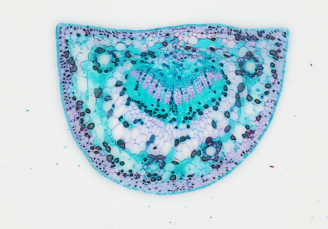
This is a cross section of a pine needle stained with safranin and fast green. Pine needles are leaves built to save water in cold or dry conditions, and this slide shows many of those adaptations.
This cross section of a pine needle (Pinus leaf c.s.) demonstrates key anatomical adaptations characteristic of xerophytic gymnosperm foliage designed to minimize transpiration while maximizing photosynthetic efficiency. The outermost layer is enclosed by a thick cutinized cuticle covering a heavily sclerified epidermis and underlying hypodermis, which provides structural integrity and prevents water loss. Sunken stomata are embedded along the margin to regulate gas exchange under arid or freezing conditions. Internally, the mesophyll consists of distinctive plicate (infolded) parenchyma cells, increasing internal surface area for chloroplast distribution. Resin ducts surrounded by specialized epithelial secretory cells run longitudinally through the mesophyll. Enclosed by a prominent endodermis with a Casparian strip, the central vascular bundle contains transfusion tissue surrounding parallel xylem and phloem strands, facilitating water transport and nutrient trans-location.
What to look for
- Cuticle and epidermis: A thick waxy coat over a layer of thick-walled cells.
- Hypodermis: Thick-walled, often red-stained cells just under the epidermis that add strength.
- Sunken stomata: Stomata set down into the surface, which reduces water loss.
- Mesophyll: Photosynthetic cells with folded walls that increase surface area.
- Resin ducts: Round canals lined with small cells, spaced through the mesophyll.
- Endodermis and vascular tissue: A ring of cells around the central vascular region, with xylem stained red.
At each magnification
Low power (about 4x)
A half-moon or triangular section with a central core and small round canals.
Medium power (about 10x)
Resin ducts, the ring of endodermis, and the vascular tissue in the center.
High power (about 40x)
The folded walls of mesophyll cells and the sunken stomata.
Questions and answers
What are the parts of a pine needle cross section?
A pine needle has a thick cuticle, epidermis, and hypodermis on the outside, sunken stomata, mesophyll cells with folded walls, resin ducts, and a central vascular region surrounded by an endodermis.
How are pine needles adapted to save water?
Pine needles have a small surface area, a thick waxy cuticle, a tough layer of thick-walled cells under the skin, and stomata sunk below the surface. Together, these reduce water loss in dry or freezing weather.
- Category
- Botany & Plants
- Scan magnification
- 20x
- Stain
- Safranin and Fast Green
- Tags
- pine needle cross section, Pinus, gymnosperm anatomy, pine leaf c.s., plicate mesophyll, resin duct
Viewing this slide in class
This is a scan of a real prepared slide, not a simulation or a drawing. On StudyScope you can pan from the whole slide down to individual structures, always in focus, and measure them in real micrometers with the calibrated ruler. Present it live and every student screen follows your view and annotations. Students join with a room code on Chromebooks, iPads, or any browser, with no accounts.