Pumpkin Stem Cross Section Under the Microscope
Pumpkin Stem (Cross Section), Cucurbita pepo
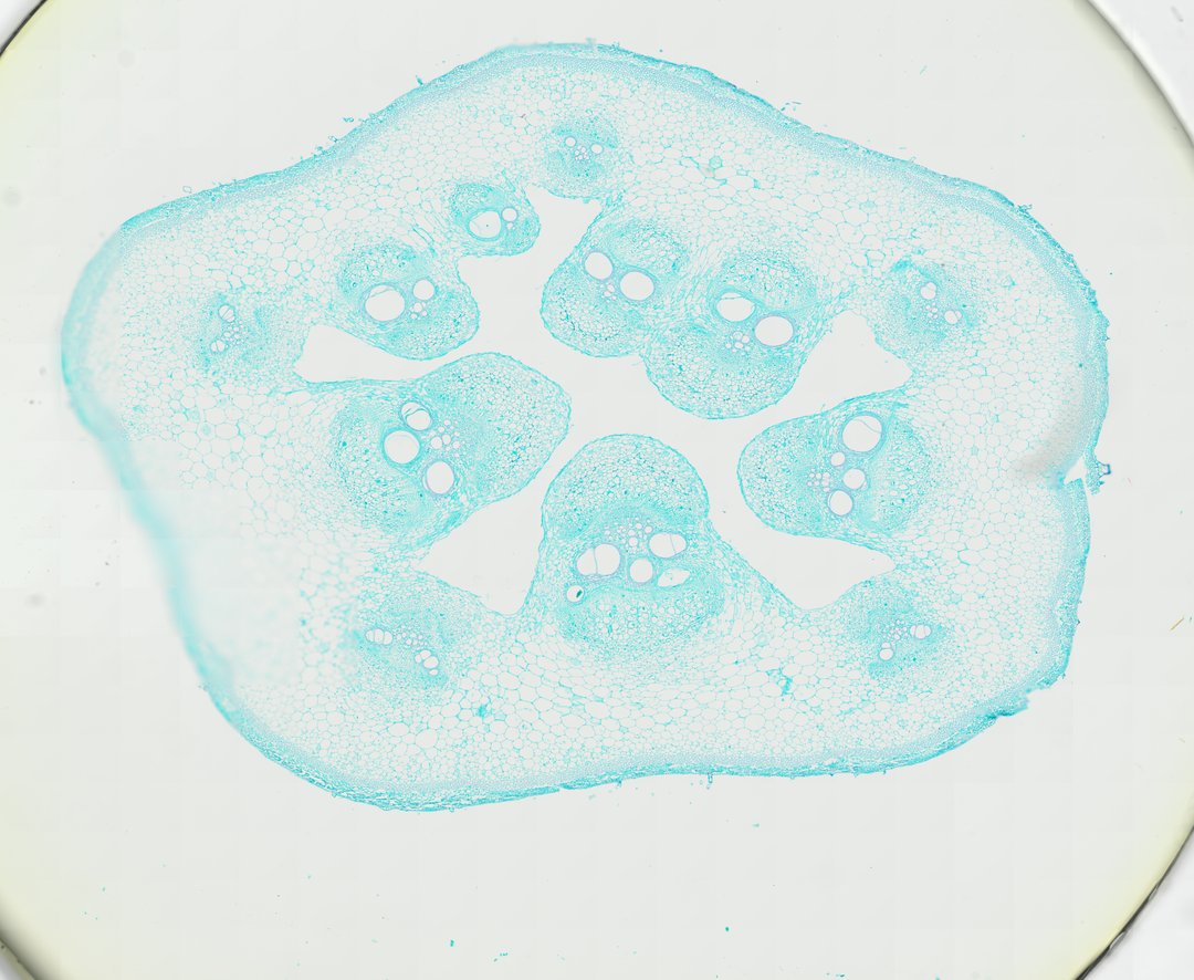
This is a cross section of a pumpkin stem stained with fast green. Pumpkin stems have unusual vascular bundles with phloem on both sides of the xylem, making it a popular slide for studying plant transport tissues.
A cross section of a pumpkin stem (Cucurbita pepo stem c.s.) stained with Fast Green, demonstrating the classic anatomical structure of a dicotyledonous herbaceous vine. The stem features a prominent pentagonal arrangement of open bicollateral vascular bundles arranged around a hollowed central pith region. Each bicollateral bundle contains large, thick-walled xylem vessels flanked by outer and inner phloem tissue separated by fascicular cambium. The prominent outer cortex consists of collenchyma and parenchyma cells providing mechanical support, bounded by a continuous epidermis.
What to look for
- Epidermis: The outer layer of cells around the ridged edge of the stem.
- Cortex: Supporting and storage cells under the epidermis, including thick-cornered collenchyma.
- Vascular bundles: Large bundles arranged in rings around a hollow or loose center.
- Xylem vessels: Very large, open, round vessels in the middle of each bundle.
- Outer and inner phloem: Phloem on both the outer and inner sides of the xylem in each bundle.
- Sieve tubes: Large phloem cells in which sieve plates can sometimes be seen.
At each magnification
Low power (about 4x)
A ridged, round outline with large bundles arranged around a hollow center.
Medium power (about 10x)
Each bundle with large xylem vessels and phloem above and below.
High power (about 40x)
Sieve tubes in the phloem and the thick walls of xylem vessels.
Questions and answers
What is a bicollateral vascular bundle?
A bicollateral bundle has phloem on both sides of the xylem, one on the outside and one on the inside. Pumpkin and other squash stems are the classic example.
Why use a pumpkin stem to study phloem?
Pumpkin phloem has very large sieve tubes, so the sieve plates between them are easier to see than in most plants. The xylem vessels are also large and easy to spot.
- Category
- Botany & Plants
- Scan magnification
- 20x
- Stain
- Fast Green
- Tags
- pumpkin stem, Cucurbita pepo, pumpkin stem cross section, bicollateral vascular bundle, xylem vessel, phloem
Viewing this slide in class
This is a scan of a real prepared slide, not a simulation or a drawing. On StudyScope you can pan from the whole slide down to individual structures, always in focus, and measure them in real micrometers with the calibrated ruler. Present it live and every student screen follows your view and annotations. Students join with a room code on Chromebooks, iPads, or any browser, with no accounts.