Radish Root Cross Section Under the Microscope
Radish Root (Cross Section), Raphanus sativus
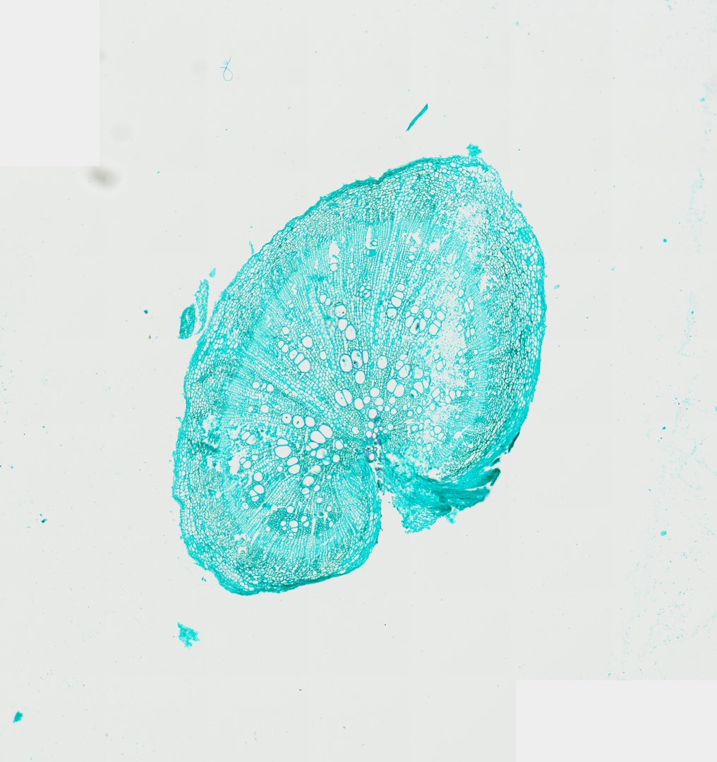
This is a cross section of a radish root stained with fast green. It shows how a storage root gets thick by adding layers of tissue full of storage cells.
This cross section of a radish (Raphanus sativus) taproot demonstrates substantial secondary growth characteristic of storage roots in Brassicaceae. The bulk of the root volume is composed of extensive secondary xylem with abundant parenchymatous storage tissue intermingled with prominent, thick-walled xylem vessels. Surrounding the vascular cylinder, a continuous vascular cambium layer gives rise to outer secondary phloem and periderm. Stained with Fast Green, the delicate cellulosic primary cell walls of the storage parenchyma and ground tissue are prominently highlighted in bright turquoise, contrasting with the structural vessel elements radiating outward from the central primary core.
What to look for
- Periderm: The protective outer skin layer of the root.
- Secondary phloem: A band of tissue just inside the outer layer.
- Vascular cambium: A thin ring of dividing cells between the phloem and the xylem.
- Secondary xylem with storage parenchyma: The large central region, mostly thin-walled storage cells, with scattered xylem vessels.
- Xylem vessels: Thick-walled, round vessels scattered in rows through the storage tissue.
At each magnification
Low power (about 4x)
A large round section, mostly storage tissue, with a thin outer rim.
Medium power (about 10x)
Rows of xylem vessels running outward from the center through the storage cells.
High power (about 40x)
Thin-walled storage cells and the thicker walls of the vessels.
Questions and answers
What does a radish root look like under a microscope?
A radish root cross section is mostly large, thin-walled storage cells, with xylem vessels scattered through them in lines from the center outward. A thin ring of cambium and phloem sits near the outside, under the skin.
Where does a radish store food?
Radishes store food in parenchyma cells mixed in with the xylem. The cambium keeps adding new cells, which is how the root swells.
- Category
- Botany & Plants
- Scan magnification
- 20x
- Stain
- Fast Green
- Tags
- radish root cross section, Raphanus sativus, radish root, taproot cross section, secondary xylem, secondary phloem
Viewing this slide in class
This is a scan of a real prepared slide, not a simulation or a drawing. On StudyScope you can pan from the whole slide down to individual structures, always in focus, and measure them in real micrometers with the calibrated ruler. Present it live and every student screen follows your view and annotations. Students join with a room code on Chromebooks, iPads, or any browser, with no accounts.