Sunflower Leaf Cross Section Under the Microscope
Sunflower Leaf (Cross Section), Helianthus annuus
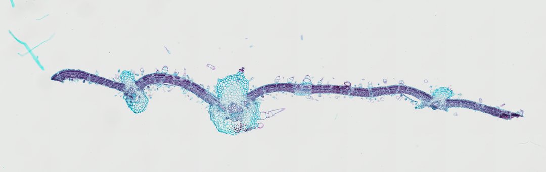
This is a cross section of a sunflower leaf stained with safranin and fast green. It shows the standard dicot leaf layers plus lots of hairs on the surfaces.
A cross section of a sunflower leaf (Helianthus annuus C.S.) double-stained with Safranin and Fast Green, illustrating the dorsiventral structure of a herbaceous dicot leaf. Numerous prominent multicellular non-glandular trichomes (hairs) project from both the upper and lower single-layered epidermal surfaces. The midrib features a large primary vascular bundle surrounded by collenchymatous supporting tissue beneath the epidermis. Lignified xylem tracheary elements stained reddish-pink face the upper adaxial side, while phloem tissue faces the abaxial side. The leaf lamina exhibits a distinct differentiation between densely packed adaxial palisade mesophyll cells and loosely arranged abaxial spongy mesophyll cells separated by intercellular air spaces.
What to look for
- Trichomes (hairs): Hairs made of several cells sticking out from the upper and lower surfaces.
- Epidermis: Single layers of cells on the top and bottom.
- Palisade mesophyll: Tall, tightly packed cells under the upper epidermis.
- Spongy mesophyll: Loosely arranged cells with air spaces toward the lower surface.
- Midrib: A large central vein with supporting tissue, red-stained xylem on top, and green-stained phloem below.
At each magnification
Low power (about 4x)
The thin leaf blade, the bulging midrib, and hairs along the edges.
Medium power (about 10x)
The palisade and spongy layers and the vascular bundle in the midrib.
High power (about 40x)
The cells that make up each hair and the red walls of the xylem.
Questions and answers
Why are sunflower leaves hairy?
The hairs, called trichomes, can slow water loss, shade the leaf surface, and make the leaf harder for insects to eat.
What are the parts of a sunflower leaf cross section?
A sunflower leaf has an upper and lower epidermis with hairs, a palisade layer of tall cells, a spongy layer with air spaces, and a midrib with a large vascular bundle, xylem on top and phloem below.
- Category
- Botany & Plants
- Scan magnification
- 20x
- Stain
- Safranin / Fast Green
- Tags
- sunflower leaf, Helianthus annuus, sunflower leaf cross section, dorsiventral leaf, leaf cross section, trichomes
Viewing this slide in class
This is a scan of a real prepared slide, not a simulation or a drawing. On StudyScope you can pan from the whole slide down to individual structures, always in focus, and measure them in real micrometers with the calibrated ruler. Present it live and every student screen follows your view and annotations. Students join with a room code on Chromebooks, iPads, or any browser, with no accounts.