Sunflower Stem Cross Section Under the Microscope
Sunflower Stem (Cross Section), Helianthus annuus
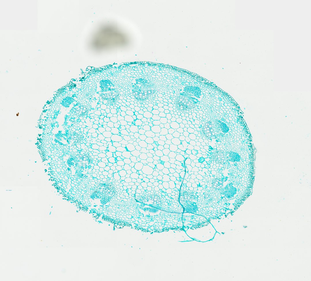
This is a cross section of a young sunflower stem stained with fast green. It is the textbook example of a dicot stem, with vascular bundles arranged in a ring around a central pith.
A cross section of a young sunflower stem (Helianthus stem c.s.) stained with Fast Green, illustrating the textbook eustele organization typical of herbaceous dicotyledonous stems. The section shows discrete collateral vascular bundles arranged in a distinct ring around a expansive central pith composed of large parenchymal cells. Each bundle features inwardly directed primary xylem vessels, an active fascicular cambium layer, and outer phloem tissue capped by prominent sclerenchyma bundle caps (fibers). Surrounding the vascular cylinder is a narrow cortex composed of collenchyma and parenchyma cells, enclosed by a single layer of epidermal cells.
What to look for
- Epidermis: A single outer layer of cells.
- Cortex: A narrow zone of supporting and storage cells under the epidermis, including thick-cornered collenchyma.
- Vascular bundles: Wedge-shaped bundles arranged in a ring.
- Xylem, cambium, and phloem: Xylem on the inner side of each bundle, phloem on the outer side, and a thin cambium between them.
- Bundle caps: Clusters of thick-walled fiber cells capping the outer side of each bundle.
- Pith: Large, thin-walled cells filling the center of the stem.
At each magnification
Low power (about 4x)
A round section with a ring of bundles around a large center.
Medium power (about 10x)
Each bundle with its fiber cap, phloem, cambium, and xylem.
High power (about 40x)
The thick walls of the xylem vessels and the small, flat cells of the cambium.
Questions and answers
What does a dicot stem cross section look like under a microscope?
A dicot stem like sunflower has an epidermis, a cortex, a ring of vascular bundles, and a central pith. In each bundle, xylem faces the center, phloem faces outward, and a thin cambium lies between them.
How is a sunflower stem different from a corn stem?
A sunflower stem is a dicot stem, with its vascular bundles in one ring around a pith. A corn stem is a monocot stem, with its bundles scattered throughout.
- Category
- Botany & Plants
- Scan magnification
- 20x
- Stain
- Fast Green
- Tags
- sunflower stem, Helianthus annuus, sunflower stem cross section, eustele, dicot stem, collateral vascular bundle
Viewing this slide in class
This is a scan of a real prepared slide, not a simulation or a drawing. On StudyScope you can pan from the whole slide down to individual structures, always in focus, and measure them in real micrometers with the calibrated ruler. Present it live and every student screen follows your view and annotations. Students join with a room code on Chromebooks, iPads, or any browser, with no accounts.