Wheat Stem Cross Section Under the Microscope
Wheat Stem (Cross Section), Triticum aestivum
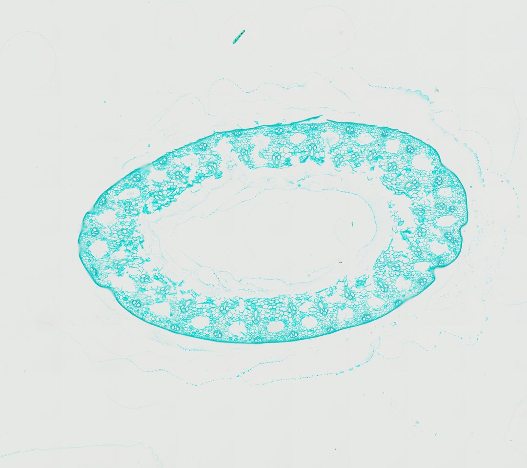
This is a cross section of a wheat stem stained with fast green. It is a classic grass stem, hollow in the middle with vascular bundles arranged in two rings.
A cross section of a wheat stem (Triticum aestivum stem c.s.) stained with Fast Green, revealing the classic architectural features of a hollow cereal grass culm. The tissue displays a hollow central pith cavity (lacuna) surrounded by a ring of ground parenchyma tissue containing closed collateral vascular bundles arranged in two distinct concentric rings. Smaller vascular bundles reside near the outer subepidermal sclerenchyma layer, while larger bundles are situated deeper within the ground tissue. Each vascular bundle contains typical monocot xylem elements, including distinct metaxylem vessels and protoxylem lacunae, paired with outer phloem tissue and encircled by a structural sclerenchymatous bundle sheath.
What to look for
- Hollow center: A large empty space in the middle of the stem.
- Epidermis: The outer layer of small cells.
- Sclerenchyma: Thick-walled strengthening cells just under the epidermis.
- Outer ring of bundles: Small vascular bundles near the outside of the stem.
- Inner ring of bundles: Larger vascular bundles deeper in the ground tissue.
- Bundle sheath: A ring of thick-walled cells around each bundle.
At each magnification
Low power (about 4x)
A ring of tissue around a hollow center.
Medium power (about 10x)
Two rings of bundles, small ones on the outside and larger ones inside.
High power (about 40x)
The large xylem vessels in each bundle and the thick-walled sheath around it.
Questions and answers
Why are wheat stems hollow?
A hollow tube is strong for its weight. Grass stems like wheat use a hollow center with a strong outer ring of tissue, which lets them stand tall and bend in the wind without using much material.
What does a monocot grass stem look like under a microscope?
A grass stem like wheat is a hollow tube with vascular bundles in the ground tissue around the center. Each bundle has large xylem vessels, phloem, and a sheath of thick-walled cells.
- Category
- Botany & Plants
- Scan magnification
- 20x
- Stain
- Fast Green
- Tags
- wheat stem, Triticum aestivum, wheat stem cross section, monocot stem, poaceae, culm
Viewing this slide in class
This is a scan of a real prepared slide, not a simulation or a drawing. On StudyScope you can pan from the whole slide down to individual structures, always in focus, and measure them in real micrometers with the calibrated ruler. Present it live and every student screen follows your view and annotations. Students join with a room code on Chromebooks, iPads, or any browser, with no accounts.