Bird Feather Under the Microscope
Bird Feather (Whole Mount), Aves
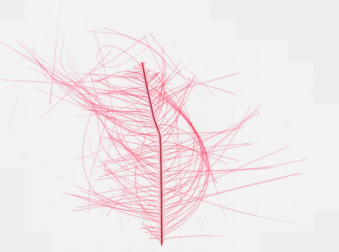
This slide shows part of a bird feather mounted whole and stained red. Students use it to see how feathers branch and hook together to form a smooth, strong surface.
This whole mount slide preparation demonstrates the microstructural architecture of a bird feather (class Aves). Stained red for enhanced visual contrast under brightfield illumination, the specimen highlights the central longitudinal shaft (rachis) from which parallel branches (barbs) extend laterally. Extending outward from the barbs are microscopic barbules, some bearing tiny interlocking hooklets (barbicels) that demonstrate how plumulaceous or pennaceous feather vanes maintain structural integrity or soft insulation. Feather keratin structures such as these are essential for avian flight, thermoregulation, and water repellency, providing a classic example of integumentary evolution in vertebrates.
What to look for
- Rachis: The thick central shaft running down the middle of the feather.
- Barbs: Thin branches coming off both sides of the rachis, lined up in parallel rows.
- Barbules: Even smaller branches coming off each barb, giving a fuzzy or fringed look.
- Hooklets (barbicels): Tiny hooks on some barbules that catch neighboring barbules, like Velcro.
At each magnification
Low power (about 4x)
The central shaft with rows of barbs branching off it.
Medium power (about 10x)
Barbules along each barb, forming a fine fringe.
High power (about 40x)
The tiny hooklets on barbules where neighboring barbs lock together.
Questions and answers
What are the parts of a feather under a microscope?
A feather has a central shaft called the rachis, branches called barbs, and smaller branches off the barbs called barbules. Many barbules have tiny hooks that link them together.
How do feathers stay together?
Barbules on one barb have tiny hooks that catch onto barbules of the next barb, zipping them together. When birds preen, they are re-hooking barbs that have pulled apart.
What are feathers made of?
Feathers are made of keratin, the same kind of protein found in hair, nails, and scales.
- Category
- Vertebrates
- Scan magnification
- 20x
- Stain
- Red stain
- Tags
- bird feather, Aves, feather whole mount, rachis, barb, barbule
Viewing this slide in class
This is a scan of a real prepared slide, not a simulation or a drawing. On StudyScope you can pan from the whole slide down to individual structures, always in focus, and measure them in real micrometers with the calibrated ruler. Present it live and every student screen follows your view and annotations. Students join with a room code on Chromebooks, iPads, or any browser, with no accounts.