Fish Gill Cross Section Under the Microscope
Fish Gill (Cross Section), Actinopterygii
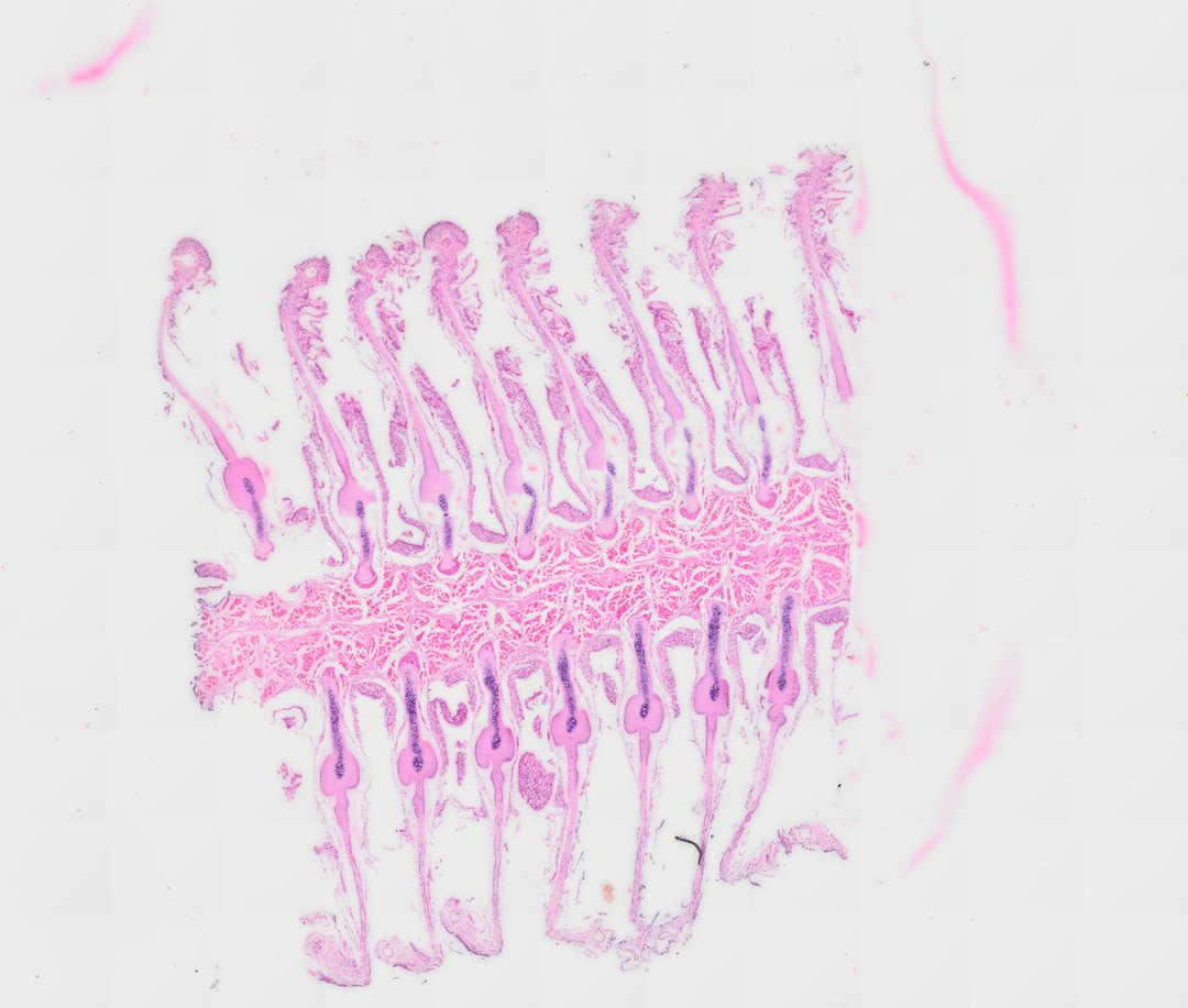
This is a section through a fish gill stained with H&E. It shows how fish get oxygen from water using thin, feathery folds packed with blood vessels.
This cross section of a teleost fish gill demonstrates the structural architecture essential for respiratory gas exchange in aquatic environments. The slide reveals the central bony or cartilaginous gill arch supporting double rows of primary gill filaments (primary lamellae). Extending laterally from each primary filament are delicate, highly vascularized secondary lamellae lined by a thin simple squamous epithelium. Hematoxylin and eosin (H&E) staining highlights the underlying cartilaginous ray support, capillary networks packed with nucleated erythrocytes, and mucous or ionoregulatory chloride cells located along the interlamellar regions.
What to look for
- Gill arch: The thick supporting bar that the gill filaments attach to.
- Gill filaments: Long, finger-like projections extending from the arch in rows.
- Secondary lamellae: Tiny, thin plates lining both sides of each filament, like the teeth of a comb.
- Blood cells: Small oval red blood cells with nuclei, stained pink to red, inside the lamellae.
- Cartilage support: A pale rod running along the inside of each filament.
At each magnification
Low power (about 4x)
Rows of long filaments branching from the gill arch.
Medium power (about 10x)
The comb-like lamellae along each filament.
High power (about 40x)
Red blood cells in the lamellae, separated from the water by a very thin layer of cells.
Questions and answers
What does a fish gill look like under a microscope?
A fish gill has a supporting arch with rows of long filaments. Each filament is lined with many thin plates called lamellae, which are packed with tiny blood vessels, giving the gill a feathery look.
How do gills get oxygen from water?
Water flows over the thin lamellae while blood flows through them in the opposite direction. This countercurrent flow keeps oxygen moving from the water into the blood along the whole length of the lamella.
Why are gill lamellae so thin?
Oxygen passes into the blood by diffusion, which works best over short distances. Thin lamellae put the blood very close to the water and give a huge surface area for gas exchange.
- Category
- Vertebrates
- Scan magnification
- 20x
- Stain
- H&E
- Tags
- fish gill, gill filaments, primary lamellae, secondary lamellae, gill arch, Actinopterygii
Viewing this slide in class
This is a scan of a real prepared slide, not a simulation or a drawing. On StudyScope you can pan from the whole slide down to individual structures, always in focus, and measure them in real micrometers with the calibrated ruler. Present it live and every student screen follows your view and annotations. Students join with a room code on Chromebooks, iPads, or any browser, with no accounts.