Frog Epidermic Cell Cross Section Under the Microscope
Frog Epidermic Cell (Cross Section), Rana temporaria
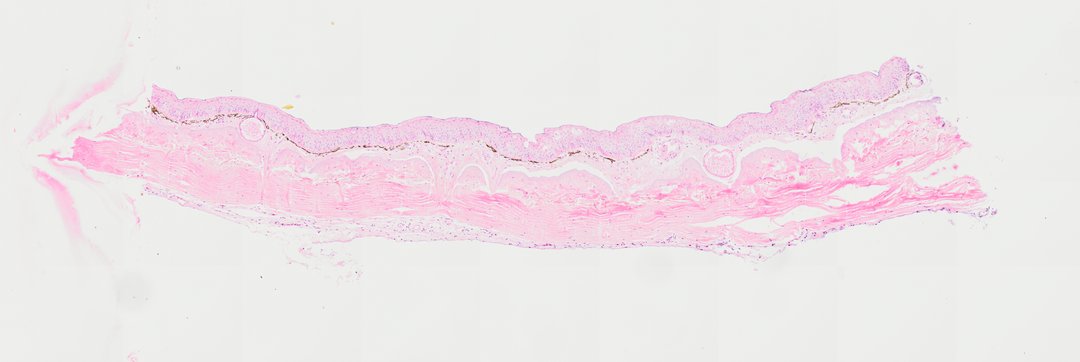
This is a cross section of frog skin stained with H&E. Frog skin is thin, moist, and full of glands, and frogs even use it to breathe, so it makes a good comparison with human skin.
A cross section (Section) of frog skin highlighting epidermal structures (labeled Frog Epidermic Cell Section). The slide displays a stratified squamous epithelium forming the epidermis, underlain by a dermis containing cutaneous glands (mucous and poison glands) and pigment-containing chromatophores, demonstrating typical amphibian integumentary histology.
What to look for
- Epidermis: The outer layer of several rows of cells, with purple nuclei.
- Dermis: Pink connective tissue under the epidermis.
- Mucous glands: Small round glands in the dermis that keep the skin moist.
- Poison (granular) glands: Larger glands in the dermis that release protective chemicals.
- Pigment cells: Dark cells (chromatophores) near the top of the dermis that give frogs their color.
At each magnification
Low power (about 4x)
A thin strip of skin with a darker outer layer and round glands below it.
Medium power (about 10x)
The layers of the epidermis and the glands in the dermis.
High power (about 40x)
Individual cells of the epidermis and the dark pigment cells.
Questions and answers
What does frog skin look like under a microscope?
Frog skin has a thin epidermis of several cell layers on top of a dermis full of round glands. Some glands make mucus to keep the skin moist, and others make chemicals that protect the frog. Dark pigment cells give the skin its color.
Why is frog skin always moist?
Mucous glands keep frog skin wet. Frogs take in some of their oxygen through their skin, and gases pass through a moist surface much more easily.
- Category
- Vertebrates
- Scan magnification
- 20x
- Stain
- H&E
- Tags
- Frog Epidermic Cell Section, Frog epidermic cell section, Frog skin section, Frog skin cross section, Frog skin, Frog epidermis
Viewing this slide in class
This is a scan of a real prepared slide, not a simulation or a drawing. On StudyScope you can pan from the whole slide down to individual structures, always in focus, and measure them in real micrometers with the calibrated ruler. Present it live and every student screen follows your view and annotations. Students join with a room code on Chromebooks, iPads, or any browser, with no accounts.