Ovary of Fish Under the Microscope
Ovary of Fish (Section), Cyprinus carpio
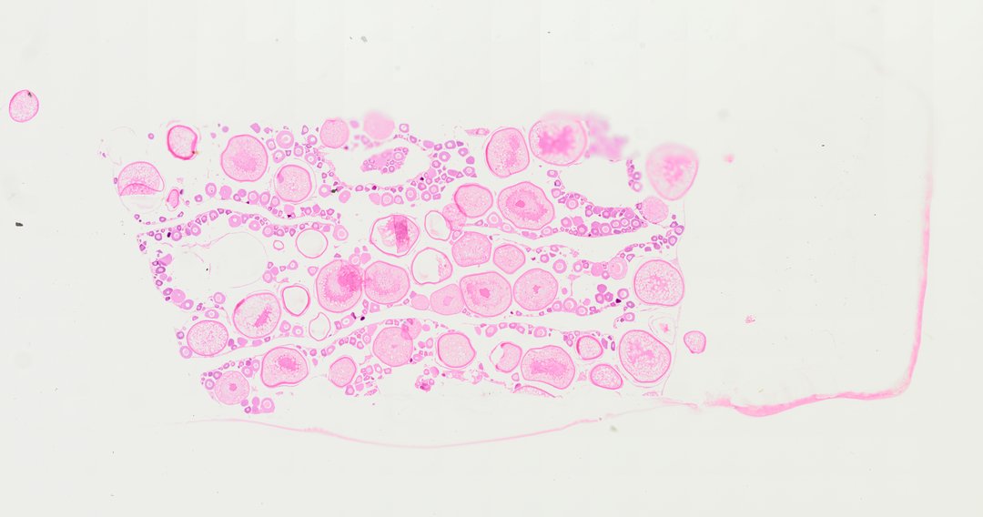
This is a section of a fish ovary stained with H&E. It shows egg cells (oocytes) at many sizes, from small young ones to large ones packed with yolk.
A sectional view (Sec.) of a fish ovary (labeled Ovary of Fish Sec.). The slide displays mature and developing oocytes filled with yolk granules, surrounded by follicular layers within the ovarian lumen, illustrating teleost female reproductive histology.
What to look for
- Young oocytes: Small cells with a large nucleus and cytoplasm that stains purple.
- Mature oocytes: Large round cells filled with pink-staining yolk.
- Nucleus: A large round nucleus near the center of growing oocytes.
- Follicle layer: A thin layer of cells wrapped around each oocyte.
- Ovarian tissue: Thin pink connective tissue holding the oocytes in place.
At each magnification
Low power (about 4x)
Many round egg cells of very different sizes packed together.
Medium power (about 10x)
Small purple young oocytes next to large pink yolk-filled ones.
High power (about 40x)
The yolk granules and the thin follicle layer around a large oocyte.
Questions and answers
What does a fish ovary look like under a microscope?
A fish ovary is packed with round egg cells of many sizes. Small, young oocytes stain purple with H&E, while larger, older ones fill with yolk and stain pink.
Why are fish eggs so different in size on one slide?
Fish ovaries hold eggs at many stages of development at the same time. As each egg grows, it builds up yolk, which feeds the embryo after fertilization.
- Category
- Vertebrates
- Scan magnification
- 20x
- Stain
- H&E
- Tags
- Ovary of Fish Sec., Ovary of fish section, Fish ovary sec, Fish ovary section, Fish ovary cross section, Fish ovary
Viewing this slide in class
This is a scan of a real prepared slide, not a simulation or a drawing. On StudyScope you can pan from the whole slide down to individual structures, always in focus, and measure them in real micrometers with the calibrated ruler. Present it live and every student screen follows your view and annotations. Students join with a room code on Chromebooks, iPads, or any browser, with no accounts.