Frog Blood Smear Under the Microscope
Frog Blood Smear, Rana temporaria
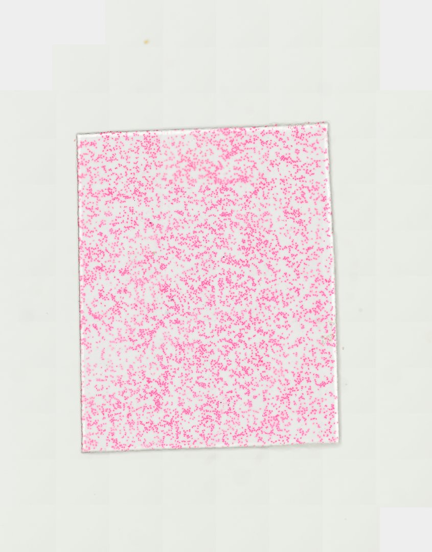
This is a smear of frog blood stained with Giemsa. Frog red blood cells are large, oval, and have a nucleus, which makes them easy to see and good for comparing with human blood.
A blood smear preparation of frog blood (labeled Frog blood smear). The slide displays large, oval, nucleated amphibian erythrocytes (red blood cells) along with various white blood cells, illustrating characteristic non-mammalian vertebrate hematology.
What to look for
- Red blood cells: Large oval cells with pinkish cytoplasm and a dark purple oval nucleus in the center.
- White blood cells: Rounder cells with larger or oddly shaped nuclei, far fewer than red cells.
- Thrombocytes: Small cells with a nucleus that help blood clot.
At each magnification
Low power (about 4x)
A thin film of cells across the slide.
Medium power (about 10x)
Many large oval red cells, each with a dark nucleus.
High power (about 40x)
Details of the nucleus and the different kinds of white blood cells.
Questions and answers
What does frog blood look like under a microscope?
Frog blood is mostly large, oval red blood cells with a dark nucleus in the middle of each one. A few white blood cells and small thrombocytes are scattered among them.
How is frog blood different from human blood?
Frog red blood cells are oval, have a nucleus, and are much larger, roughly two to three times the length of a human red blood cell. Human red blood cells are small, round, and have no nucleus.
- Category
- Vertebrates
- Scan magnification
- 20x
- Stain
- Giemsa
- Tags
- Frog blood smear, Frog blood, Frog blood cell smear, Rana temporaria, Rana, Amphibia
Viewing this slide in class
This is a scan of a real prepared slide, not a simulation or a drawing. On StudyScope you can pan from the whole slide down to individual structures, always in focus, and measure them in real micrometers with the calibrated ruler. Present it live and every student screen follows your view and annotations. Students join with a room code on Chromebooks, iPads, or any browser, with no accounts.