Onion Epidermis Under the Microscope
Onion Epidermis (Whole Mount), Allium cepa
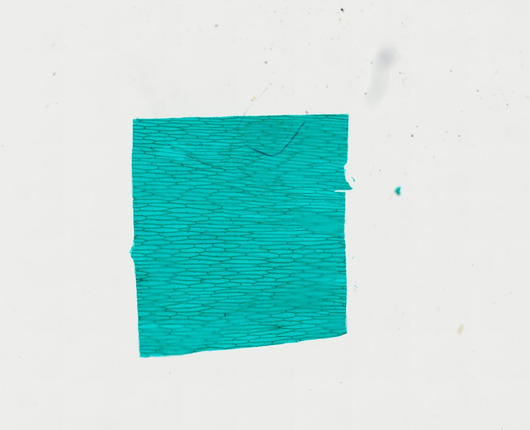
This is a thin layer of onion skin (epidermis) mounted flat and stained with fast green. It is one of the most common first slides in biology because its large, simple plant cells are easy to see.
A whole mount preparation of onion scale leaf epidermis (Allium cepa W.M.) stained with Fast Green, revealing a uniform sheet of elongated, rectangular plant epidermal cells. The single layer of cells displays distinct, thick cellulose cell walls enclosing large central vacuoles, with compressed cytoplasmic regions and subtle nuclear structures pushed against the periphery. The regular, interlocking tiling pattern of the epidermal cells serves as a classic model for studying plant tissue organization, cellular boundaries, and basic plant cytology without complex structural interference from internal mesophyll layers.
What to look for
- Cell wall: Clear green outlines around each long, rectangular cell.
- Nucleus: A small round or oval body, often pressed near the side of the cell.
- Vacuole: A large central space that fills most of each cell and pushes the cytoplasm to the edges.
- Cytoplasm: A thin layer lining the inside of the cell wall.
At each magnification
Low power (about 4x)
A sheet of long, brick-like cells lined up in rows.
Medium power (about 10x)
Individual cells with clear walls, and a nucleus in many of them.
High power (about 40x)
The nucleus and the thin layer of cytoplasm around the large vacuole.
Questions and answers
What do onion cells look like under a microscope?
Onion epidermis cells are long rectangles packed together like bricks. Each cell has a clear cell wall, a nucleus often near one side, and a large vacuole that fills most of the cell.
Why don't onion cells have chloroplasts?
The onion layers used for this slide are part of the bulb, which grows underground where there is no light. Without light for photosynthesis, these cells do not need chloroplasts.
How are onion cells different from cheek cells?
Onion cells are plant cells, so they have a rigid cell wall, a boxy shape, and a large vacuole. Cheek cells are animal cells, with no cell wall and a more irregular, flat shape.
- Category
- Botany & Plants
- Scan magnification
- 20x
- Stain
- Fast Green
- Tags
- onion epidermis, Allium cepa, onion epidermis whole mount, epidermal tissue, plant epidermis, scale leaf
Viewing this slide in class
This is a scan of a real prepared slide, not a simulation or a drawing. On StudyScope you can pan from the whole slide down to individual structures, always in focus, and measure them in real micrometers with the calibrated ruler. Present it live and every student screen follows your view and annotations. Students join with a room code on Chromebooks, iPads, or any browser, with no accounts.