Short answer: The big difference between a cheek cell and an onion cell is the cell wall. Onion epidermis cells are plant cells: long, boxy rectangles with a stiff cell wall and a large central vacuole that fills most of each cell. Cheek cells are animal cells: thin, flat and irregular, with no cell wall and no large vacuole. What they share is a nucleus, cytoplasm and a cell membrane. Neither has chloroplasts, because onion skin from a bulb does not photosynthesize.
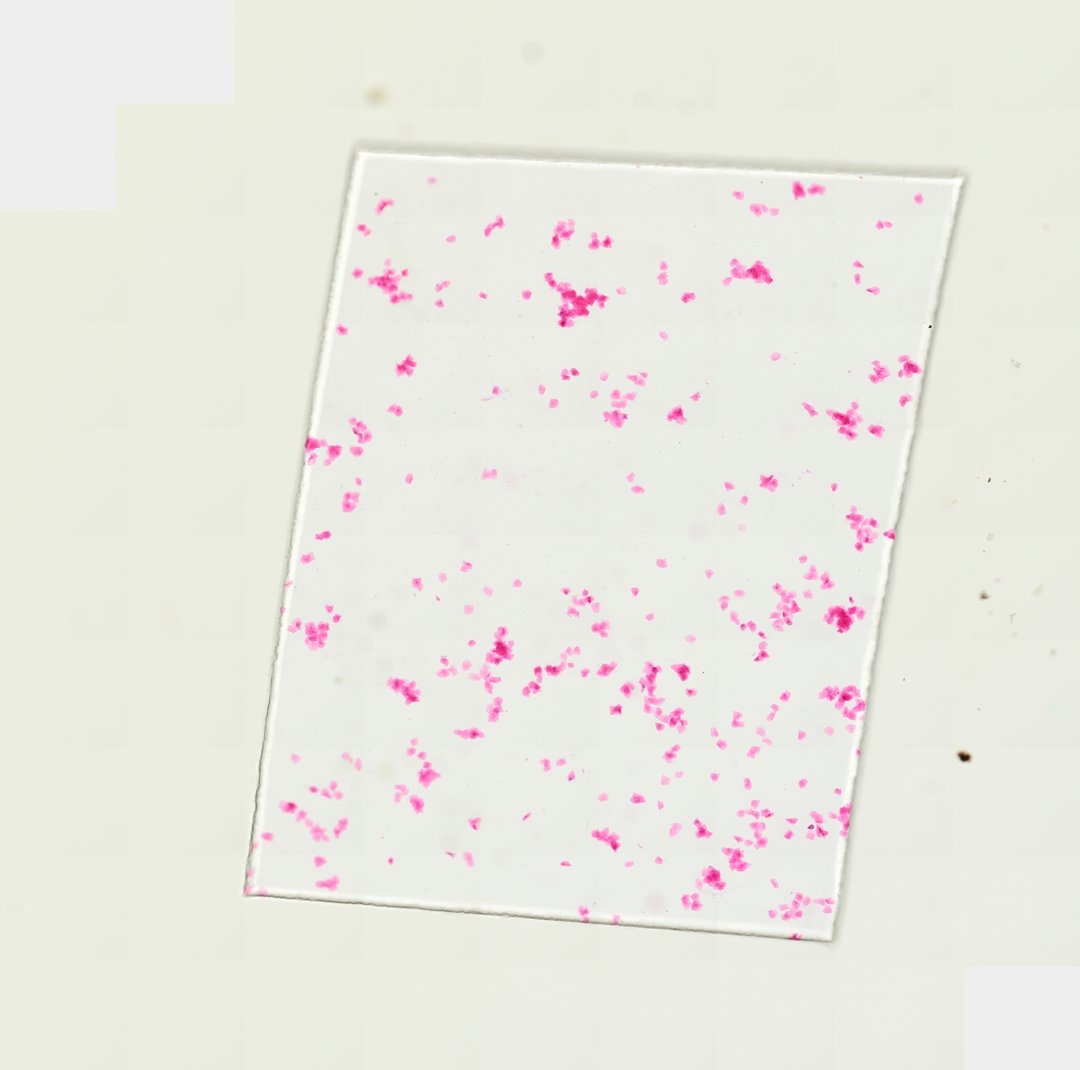
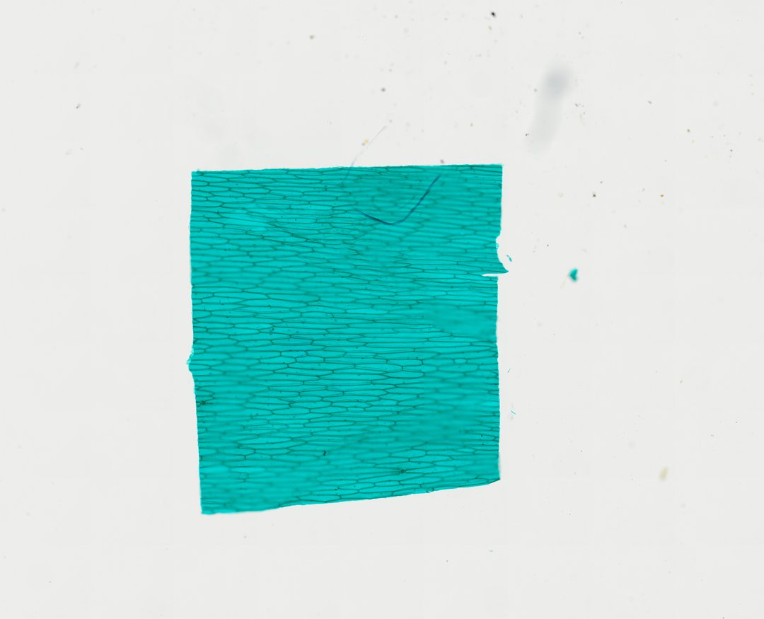
Cheek cell vs onion cell at a glance
| Feature | Cheek cell (animal) | Onion epidermis cell (plant) |
|---|---|---|
| Shape | Thin, flat and irregular, with many uneven sides | Long, narrow rectangles lined up in rows like bricks |
| Cell wall | None. Only a thin cell membrane at the edge, so cells can fold or wrinkle | Yes. A stiff cellulose wall outlines every cell and gives it a boxy shape |
| Nucleus | Small, darker, usually near the middle of the cell | Small and dark, easiest to spot in some cells, not all |
| Vacuole | No large central vacuole | One large central vacuole fills most of the cell and pushes the cytoplasm to the edges |
| Chloroplasts | None (animal cells never have them) | None in onion epidermis. This skin comes from an underground bulb, so it does not need them |
| How they sit on the slide | Scattered: some alone, some overlapping in clumps | A single sheet, one cell thick, with cells tightly packed |
| Stain on our slide | Eosin (pink) | Fast green (green walls) |
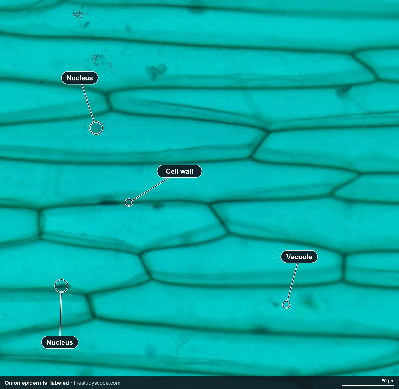
Labeled cheek cells and onion epidermis
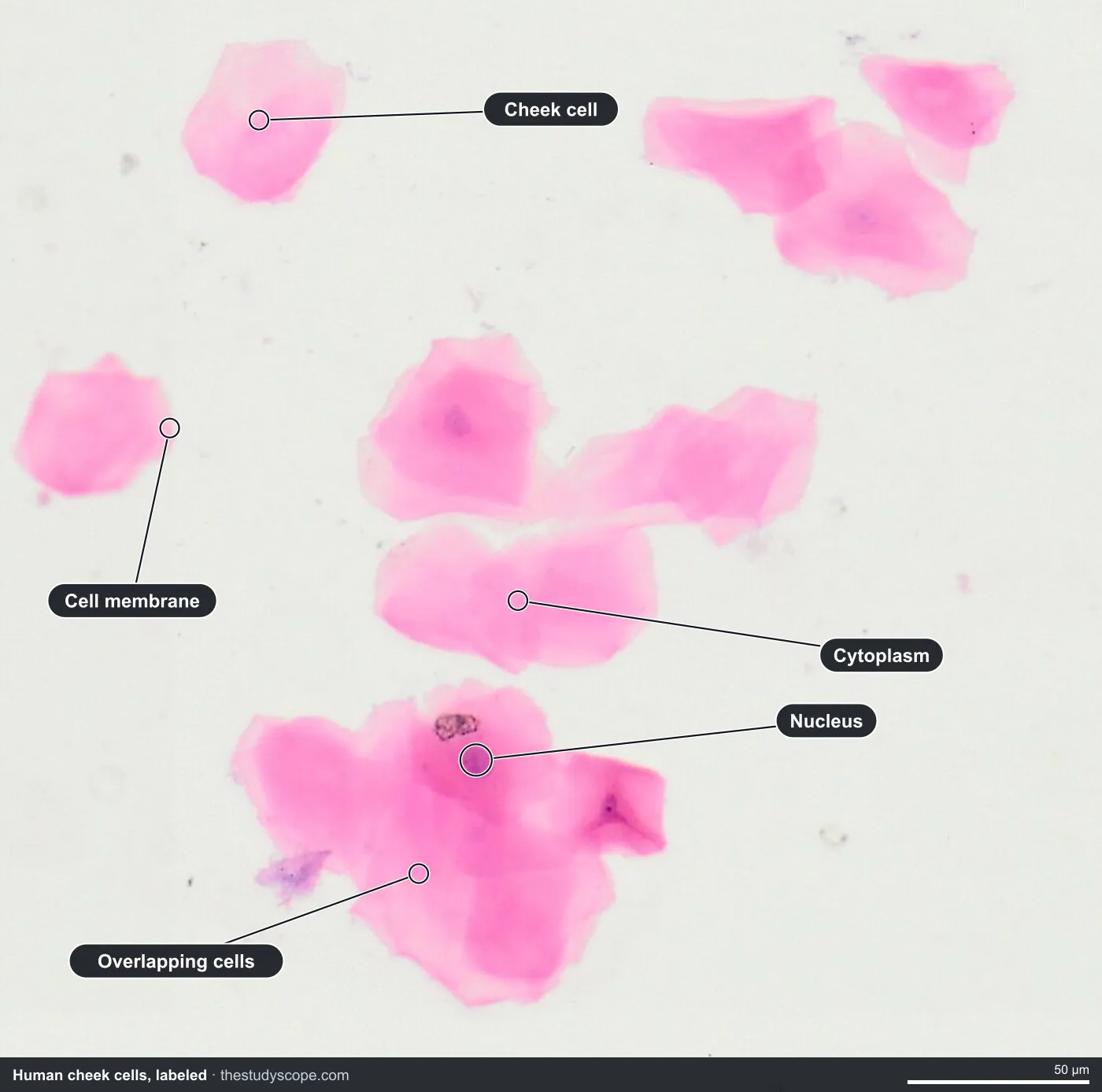
What to look for in the cheek cells
- Flat, irregular cells spread across the slide. Some lie alone, and others overlap in clumps.
- Nucleus: a small round to oval body near the center of each cell, darker than the cytoplasm around it.
- Cytoplasm: the pink-stained material filling most of each cell.
- Cell membrane: a thin outer edge with no cell wall outside it.
- Folds: thin, flexible cells can crumple, so a dark line is often a fold and not a structure.
What to look for in the onion epidermis
- Cell wall: clear green outlines around each long, rectangular cell.
- Nucleus: a small dark body, visible in some of the cells.
- Large vacuole: the big open space that fills most of each cell.
- Brick pattern: at low zoom the whole sheet looks like rows of bricks in a wall.
What cheek cells and onion cells have in common
Both are eukaryotic cells, so each has a nucleus holding its DNA, cytoplasm, and a cell membrane around the outside. Both are easy to find as a first slide because the cells are large and simple. The differences you can spot (wall, shape, vacuole) all trace back to the same idea: plants need a rigid frame and water storage, while animal cells are soft and flexible.
Measure the cells
Every slide is calibrated from its scan, so the ruler in the viewer reads real micrometers at any zoom. Have students measure the length of five cheek cells and five onion cells, then average each set. Textbooks usually describe onion epidermis cells as noticeably longer than cheek cells, but exact sizes vary, so let the class find out what its own measurements say.
A wet-mount lab and a virtual slide teach different things
If your class can do it, scraping their own cheek cells and peeling an onion for a wet mount is a great lab. It teaches technique that a virtual slide cannot: making a mount, adding stain, focusing, and handling a thin specimen. It is also messy and uneven. Some students get folded or overlapping cells, a few see almost nothing, and not every mount stains well. A prepared slide gives every student the same well-stained cells, so the class can talk about the same thing. Many teachers use both: the wet mount first, then the real slide to see what a good result looks like.
More plant epidermis to compare
Once students can tell plant from animal, other plant skins make good extensions. Both of these are whole mounts stained with fast green:
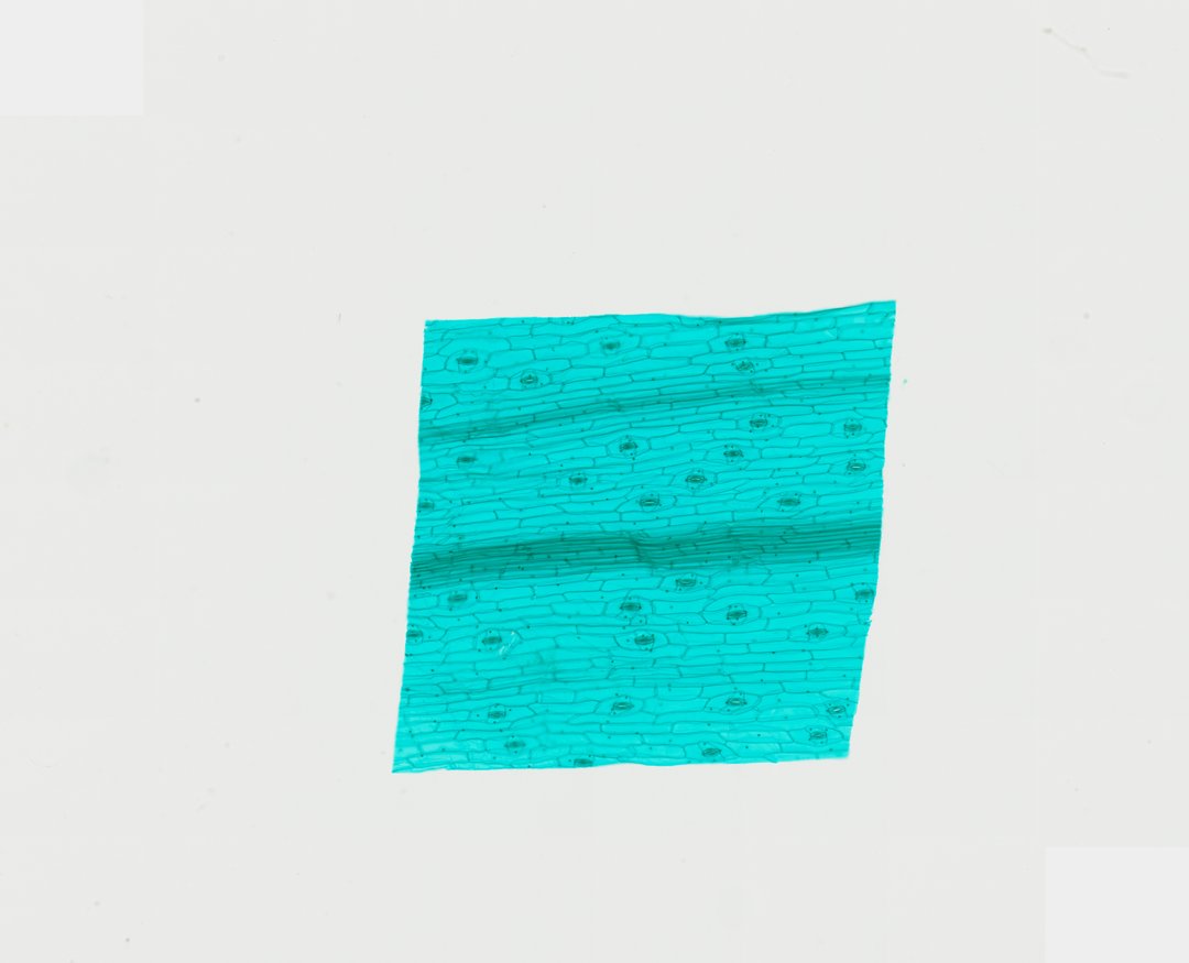
- Tradescantia (spiderwort) lower leaf epidermis: long rectangular cells in rows like onion, plus stomata, small pores with guard cells and helper cells around them.
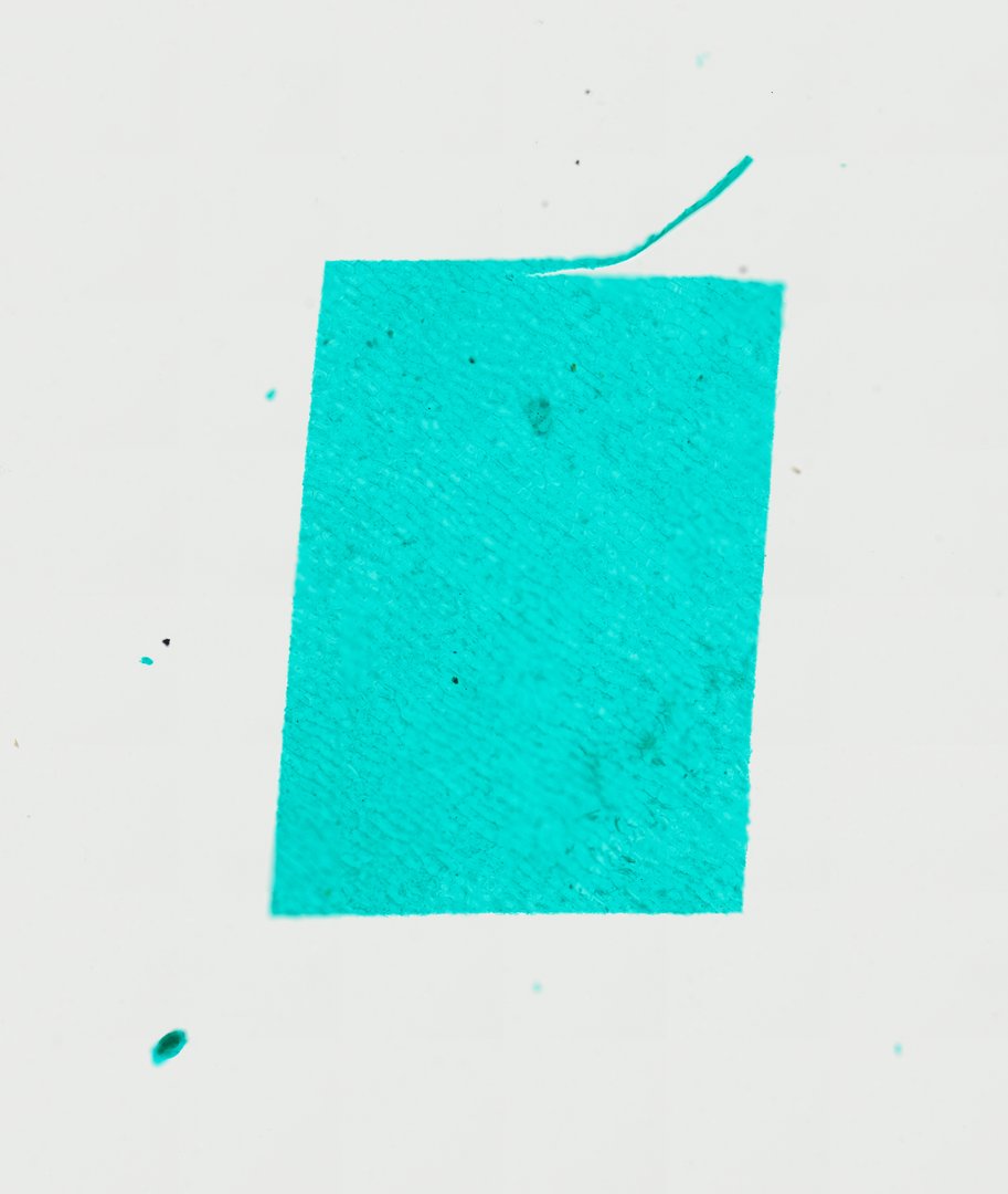
- Capsicum (pepper) peel: many-sided cells fitted together like floor tiles, a good contrast with the onion's rectangles.
Running the plant vs animal cell lab in class
- Open the cheek cells first at medium zoom and ask students to sketch one cell and list what they see.
- Open the onion epidermis at the same zoom and ask what is new. Most students spot the outlines first.
- Fill in the comparison table together: wall, shape, nucleus position, vacuole, chloroplasts.
- Measure five cells on each slide with the ruler and compare the averages.
- Have students save a stamped snapshot of their best view of each slide to keep in a notebook or slideshow.
You can present live: students join with a room code on any device, follow your view on their own screens, and see your annotations as you make them.
Ready to look? Open the cheek cells and the onion epidermis. For plant vs animal cells in a dividing context, the premade lesson Introduction to Mitosis & Cell Division covers onion root tip and roundworm eggs.
Questions and answers
What is the difference between cheek cells and onion cells?
Cheek cells are animal cells: flat and irregular, with no cell wall and no large vacuole. Onion cells are plant cells: long rectangles with a rigid cell wall and a large central vacuole. Both have a nucleus, cytoplasm and a cell membrane.
What do cheek cells and onion cells have in common?
Both are eukaryotic cells with a nucleus, cytoplasm and a cell membrane. Both are large and simple enough to see clearly at medium zoom, which is why they are the classic first slides for comparing cells.
What do cheek cells look like under a microscope?
Cheek cells are thin, flat and irregular, with a small round nucleus near the middle. Because they have no cell wall, their edges are soft and the cells can fold or overlap. On our slide they are stained pink with eosin.
What does onion skin look like under a microscope?
Onion epidermis is a sheet one cell thick. The cells are long rectangles packed in rows like bricks, each outlined by a cell wall, with a large vacuole and a small nucleus visible in some cells. On our slide the walls are stained green with fast green.
Why do onion cells have no chloroplasts?
The onion layers used for this slide come from the bulb, which grows underground without light. Without light for photosynthesis, these cells do not need chloroplasts. Plant cells in green leaves do have them.
Can students do a cheek cell and onion cell lab on a Chromebook?
Yes. StudyScope runs in any modern browser, so students can view both real prepared slides on a Chromebook, iPad or phone with nothing to install. It does not replace a wet-mount lab for learning to make a slide, but every student sees the same well-stained cells.
More guides
- Monocot vs dicot stems compared
- Frog, human, chicken and fish blood compared
- Virtual microscope, simulator or digital microscope?
- Using a microscope on a Chromebook
- How to identify the stages of mitosis in an onion root tip
- Leaf layers and stomata guide
- Pond water organisms identification guide
- How to identify tissue types
- How to measure cell size
- Lily, pine and luffa pollen compared
- Insect mouthparts compared
- Worm body cavities compared