Short answer: To measure a cell under a microscope, first find the diameter of the field of view, then divide it by the number of cells that fit across it. At low power, lay a clear ruler on the stage and read the field in millimeters, then convert to micrometers (1 mm = 1,000 micrometers). To use a higher objective, scale the field down: field at high power = field at low power x low magnification / high magnification. On a calibrated virtual slide you skip the estimating, because the ruler reads real micrometers at any zoom.
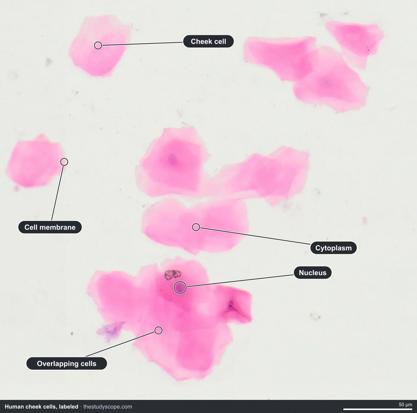
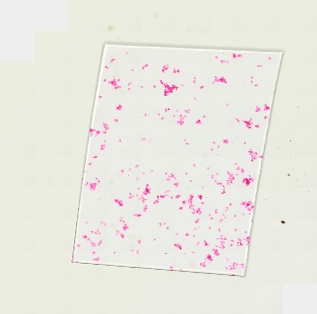 Human Cheek Cells (Whole Mount)Homo sapiensEosin
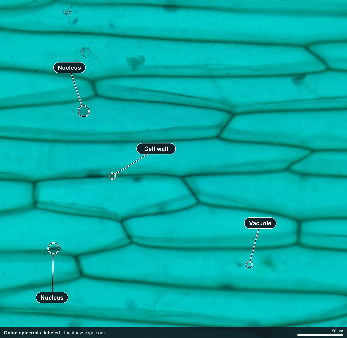
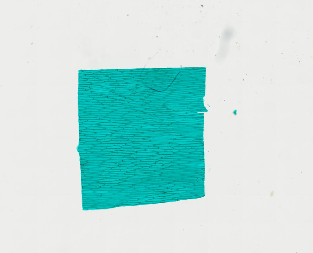 Onion Epidermis (Whole Mount)Allium cepaFast Green
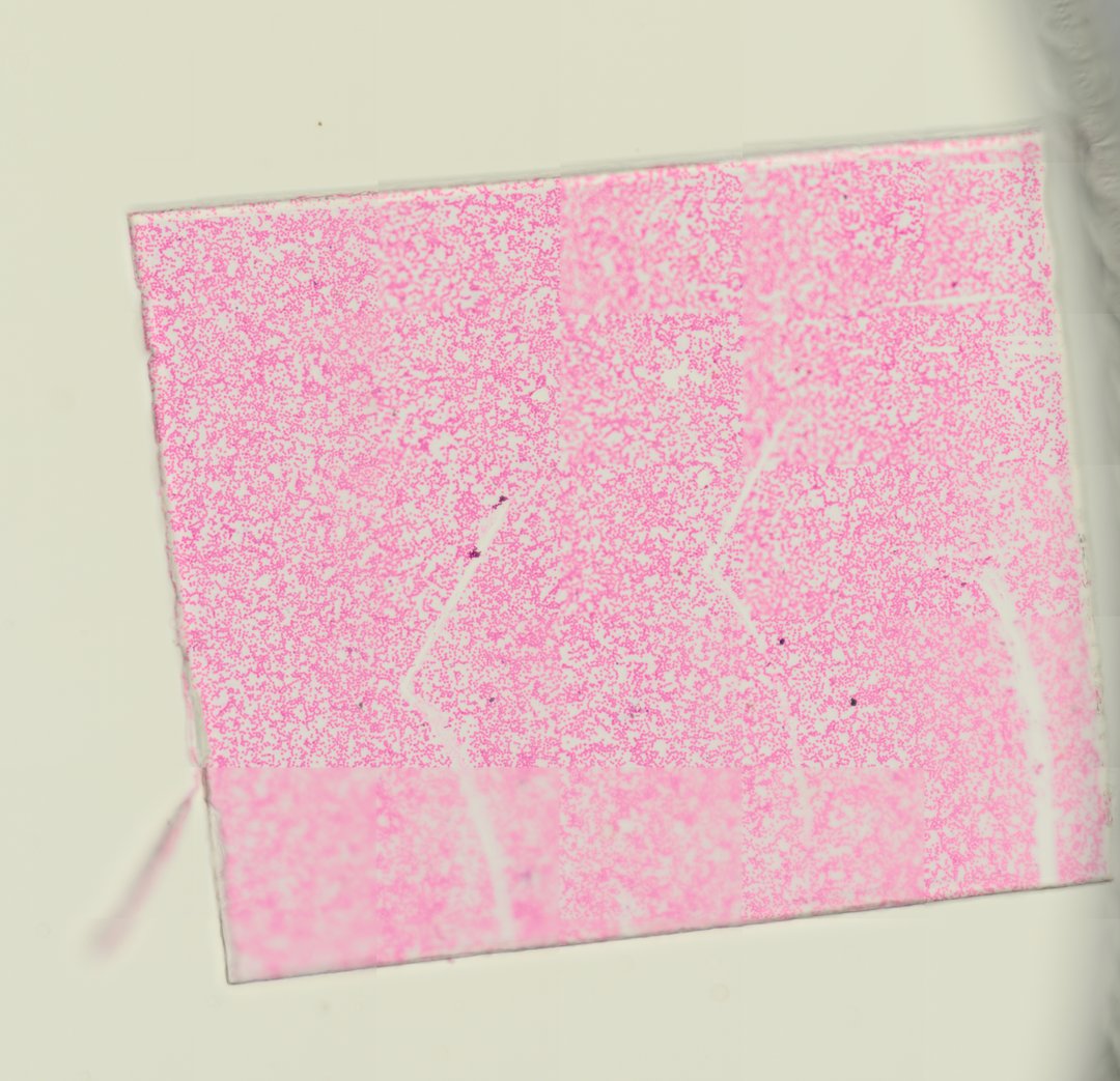 Human Blood SmearHomo sapiensWright-Giemsa
 Frog Blood SmearRana temporariaGiemsa
How to estimate cell size under a microscope: the field of view method
This is the method most textbooks teach for a bench microscope. You cannot read a cell's size straight off the eyepiece, so you measure something you can see (the circle of light called the field of view) and use it as a ruler for everything inside it.
- Measure the field of view at low power. Put the lowest objective in place and lay a clear plastic ruler (millimeter marks) across the stage opening. Line up one mark with the left edge of the circle and read how many millimeters reach the right edge.
- Convert to micrometers. Cells are too small for millimeters, so multiply by 1,000. A field of 4.5 mm is 4,500 micrometers.
- Scale the field to a higher objective. The field shrinks as magnification rises: field at high power = field at low power x low-power magnification / high-power magnification.
- Count the cells across the field at the power you are using, in a straight line from edge to edge.
- Divide. Estimated cell size = field of view diameter / number of cells across.
Worked example (illustrative numbers)
These round numbers are only an example, not a measurement from any slide. Say a ruler shows the field of view is 4.5 mm at 40x total magnification. That is 4.5 x 1,000 = 4,500 micrometers. Switching to 400x, the field is 4,500 x 40 / 400 = 450 micrometers. If 9 cells line up across that field, each cell is about 450 / 9 = 50 micrometers wide.
| Step | Calculation | Result |
|---|---|---|
| Field at 40x, in millimeters | Read from a clear ruler | 4.5 mm |
| Convert to micrometers | 4.5 x 1,000 | 4,500 micrometers |
| Field at 400x | 4,500 x 40 / 400 | 450 micrometers |
| Cells counted across at 400x | Counted by eye | 9 cells |
| Estimated cell size | 450 / 9 | About 50 micrometers |
Your estimate is only as good as your count, so use the most orderly row of cells you can find, and repeat it in two or three places. If a classroom microscope has an eyepiece reticle (a tiny ruler inside the eyepiece), you can skip the ruler on the stage: a stage micrometer (a slide with a precisely printed scale) is used once per objective to learn how many micrometers each reticle division is worth.
Total magnification: eyepiece times objective
The field of view math starts from total magnification, which is the eyepiece magnification multiplied by the objective magnification. A 10x eyepiece with a 4x objective gives 40x, and the same eyepiece with a 40x objective gives 400x.
| Objective | Eyepiece | Total magnification |
|---|---|---|
| 4x | 10x | 40x |
| 10x | 10x | 100x |
| 40x | 10x | 400x |
How to measure cells on a virtual microscope
On a StudyScope slide, the estimating steps go away. Every slide is calibrated from its scan resolution, so the ruler reads real micrometers (and millimeters) at any zoom, and the viewer shows the equivalent magnification as you zoom. Students drag across a cell and read its size instead of estimating from a count. Measuring on deep-zoom slides needs a teacher seat, and students are always free.
Estimating from the field of view is still a skill worth practicing on a real microscope, and it teaches why scale matters. For the measurement itself, the virtual ruler is faster and more precise.
Good first cells to measure
The sizes below are typical textbook ranges, so treat them as rough expectations and compare them with what your class finds.
| Cell | Typically about | Slide |
|---|---|---|
| Human red blood cell | 7 to 8 micrometers across | [Human blood smear](/catalog/slide/human-blood-smear) |
| Frog red blood cell | Roughly two to three times the length of a human red blood cell | [Frog blood smear](/catalog/slide/frog-blood-smear) |
| Human cheek cell | Several tens of micrometers across | [Oral epithelial cells](/catalog/slide/oral-epithelial-cells) |
| Onion epidermis cell | Often a few hundred micrometers long | [Onion epidermis](/catalog/slide/onion-epidermis) |
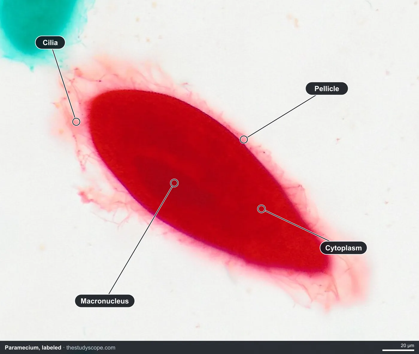
| Paramecium | Many common species are around 0.2 millimeters long | [Paramecium](/catalog/slide/paramecium) |
| Bacteria | Most are about 1 to 5 micrometers long | [Three types of bacteria smear](/catalog/slide/three-types-of-bacteria-smear) |
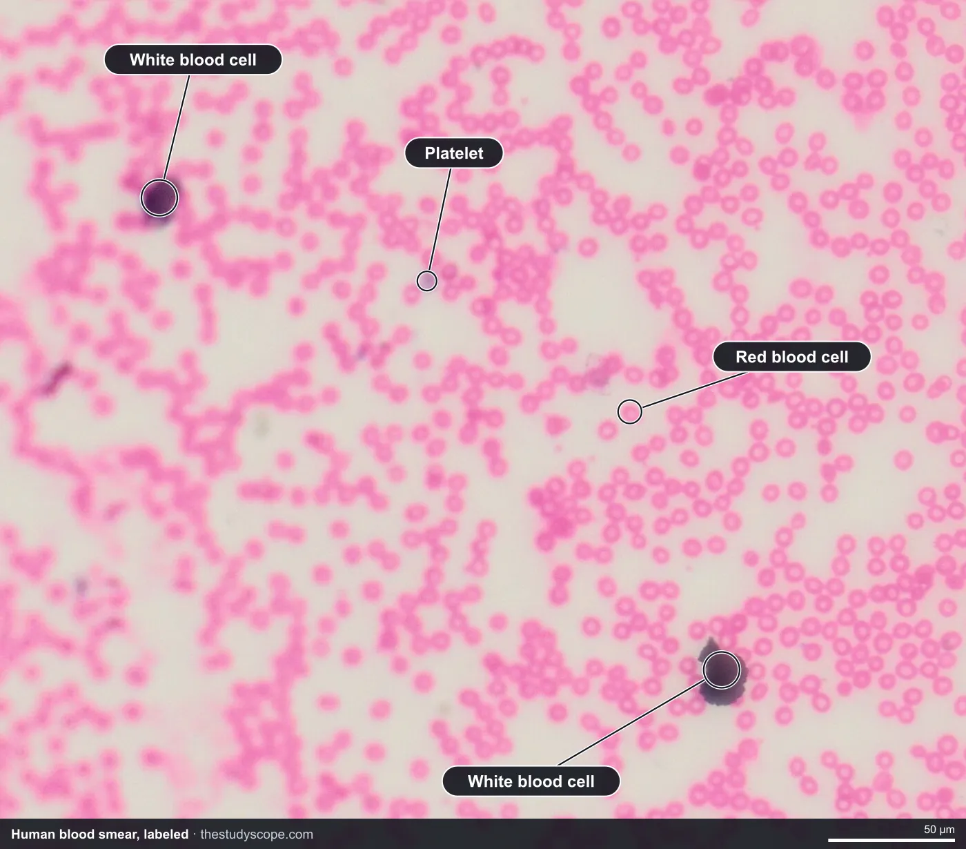
Blood cells: a built-in yardstick
Human red blood cells are a handy measuring stick because their size is so consistent. Measure a few on the human blood smear, then measure the large oval, nucleated red cells on the frog blood smear and compare. The full side-by-side is in frog vs human blood.
Cheek cells and onion cells
- On the cheek cells, measure across a flat cell, then measure its nucleus. Ask what fraction of the cell the nucleus takes up.
- On the onion epidermis, measure the length and width of one brick-shaped cell. Ask whether the length or the width is the better "size" to report.
- Compare the two: which cell is larger, and does that match what you see at the same zoom?
Paramecium and bacteria
- On the Paramecium, measure the length of one slipper-shaped cell from end to end.
- On the bacteria smear, measure the length of a short rod. Ask why you need the highest zoom to measure something this small.
- Put the sizes in order, from bacteria to Paramecium, to see the range of life that cells cover.
Teaching it
- Do the field of view calculation on paper first, using the worked example, so students know what the ruler is replacing.
- Present a slide live. Students join with a room code, with no student accounts, and follow on their own screens.
- Have each student measure three cells and compare with the typical range, then discuss why the numbers differ.
- Students can save a stamped snapshot of their view to show their measurement.
Browse the slide catalog without an account to pick your slides, starting with human blood, cheek cells and onion epidermis.
Questions and answers
How do you measure cells under a microscope?
Measure the diameter of the field of view with a clear ruler at low power, convert it to micrometers, scale it to the objective you are using, then divide by the number of cells that fit across the field. Or use an eyepiece reticle calibrated with a stage micrometer.
How do you calculate the field of view under a microscope?
Measure the field at low power with a ruler. For a higher objective, use: field at high power = field at low power x low-power magnification / high-power magnification. For example, 4.5 mm at 40x becomes 0.45 mm, or 450 micrometers, at 400x.
How do you find the diameter of a cell under a microscope?
Divide the field of view diameter by the number of cells across it. If the field is 450 micrometers and 9 cells line up edge to edge, each cell is about 50 micrometers across. This is an illustrative example.
How do you find total magnification?
Multiply the eyepiece magnification by the objective magnification. A 10x eyepiece with a 40x objective gives 400x total.
Why does the field of view get smaller at higher magnification?
Higher magnification shows a smaller piece of the slide at a larger size. Going from 40x to 400x makes the field ten times smaller across, which is why you scale it by the ratio of the two magnifications.
Can you measure cells on a virtual microscope?
Yes. Every StudyScope slide is calibrated from its scan resolution, so the ruler reads real micrometers at any zoom, and the viewer shows the equivalent magnification. Measuring needs a teacher seat, and students are always free.
More guides
- Monocot vs dicot stems compared
- Frog, human, chicken and fish blood compared
- Virtual microscope, simulator or digital microscope?
- Using a microscope on a Chromebook
- How to identify the stages of mitosis in an onion root tip
- Cheek cells vs onion cells
- Leaf layers and stomata guide
- Pond water organisms identification guide
- How to identify tissue types
- Lily, pine and luffa pollen compared
- Insect mouthparts compared
- Worm body cavities compared