Skin of Fish Cross Section Under the Microscope
Skin of Fish (Cross Section), Cyprinus carpio
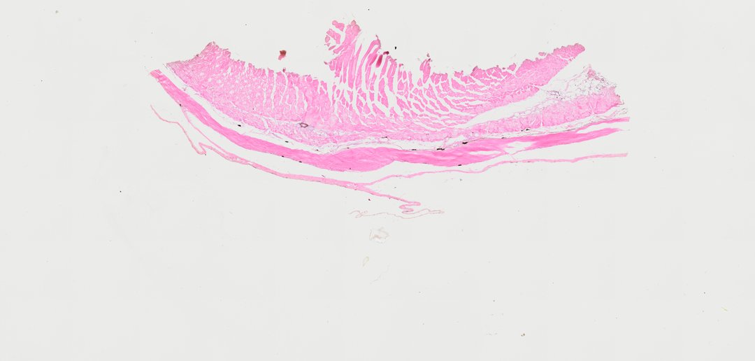
This is a section of fish skin stained with H&E. It shows that fish scales grow in the dermis and are covered by a living, mucus-making epidermis.
A sectional view (Sec.) of fish skin (labeled Skin of Fish Sec.). The slide illustrates typical teleost integumentary histology, demonstrating a multi-layered epidermis rich in mucus-secreting goblet cells, an underlying dermis containing dense connective tissue, scale pockets with developing bony scales, and deeply situated pigment cells (chromatophores).
What to look for
- Epidermis: The outer layer of several rows of cells with purple nuclei.
- Mucous (goblet) cells: Pale, rounded cells in the epidermis that make the slimy coating on fish.
- Dermis: Pink connective tissue under the epidermis.
- Scales: Thin bony plates sitting in pockets in the dermis.
- Pigment cells: Dark cells (chromatophores) in the dermis that give fish their color.
At each magnification
Low power (about 4x)
A strip of skin with the scales lying at an angle inside it.
Medium power (about 10x)
The layered epidermis over the scales and the dermis beneath.
High power (about 40x)
Mucous cells in the epidermis and dark pigment cells in the dermis.
Questions and answers
What does fish skin look like under a microscope?
Fish skin has an outer epidermis with many pale mucus-making cells, over a dermis of pink connective tissue. The scales sit in pockets in the dermis, and dark pigment cells are scattered through it.
Why are fish slimy?
Mucous cells in the epidermis make a slimy coating. It helps fish glide through water and protects them from germs and parasites.
- Category
- Vertebrates
- Scan magnification
- 20x
- Stain
- H&E
- Tags
- Skin of Fish Sec., Skin of fish section, Fish skin sec, Fish skin cross section, Fish skin section, Fish skin
Viewing this slide in class
This is a scan of a real prepared slide, not a simulation or a drawing. On StudyScope you can pan from the whole slide down to individual structures, always in focus, and measure them in real micrometers with the calibrated ruler. Present it live and every student screen follows your view and annotations. Students join with a room code on Chromebooks, iPads, or any browser, with no accounts.