Testicle of Fish Under the Microscope
Testicle of Fish (Section), Cyprinus carpio
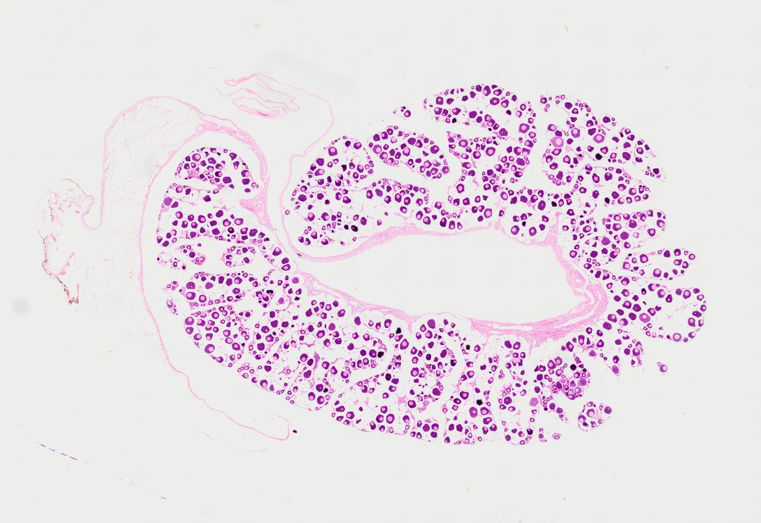
This is a section of a fish testis stained with H&E. It shows groups of cells at different stages of becoming sperm.
A sectional view (Sec.) of a fish testis (labeled Testicle of Fish Sec.). The slide shows lobular structures containing numerous spermatogenic cysts filled with developing germ cells at various stages of spermatogenesis, surrounding a central sperm duct.
What to look for
- Lobules: Compartments of the testis separated by thin pink connective tissue.
- Cysts: Groups of germ cells inside the lobules, each group at about the same stage.
- Early germ cells: Larger cells with rounder, paler nuclei.
- Late germ cells and sperm: Smaller cells with very dark, compact nuclei, often filling the center of the lobules.
- Sperm duct: A channel where mature sperm collect.
At each magnification
Low power (about 4x)
Many compartments of densely packed, purple-stained cells.
Medium power (about 10x)
Cysts with cells at different stages, some pale and some very dark.
High power (about 40x)
The size difference between large early cells and tiny, dark sperm heads.
Questions and answers
What does a fish testis look like under a microscope?
A fish testis is divided into compartments filled with groups of germ cells. Each group is at a different stage of development, from larger early cells to small, dark sperm.
Why do sperm cells stain so dark?
As sperm mature, their DNA packs very tightly into a small head. That dense DNA takes up hematoxylin strongly, so sperm heads look very dark purple.
- Category
- Vertebrates
- Scan magnification
- 20x
- Stain
- H&E
- Tags
- Testicle of Fish Sec., Testicle of fish section, Fish testicle sec, Fish testis section, Fish testis cross section, Fish testis
Viewing this slide in class
This is a scan of a real prepared slide, not a simulation or a drawing. On StudyScope you can pan from the whole slide down to individual structures, always in focus, and measure them in real micrometers with the calibrated ruler. Present it live and every student screen follows your view and annotations. Students join with a room code on Chromebooks, iPads, or any browser, with no accounts.