Ant Under the Microscope
Ant (Whole Mount), Formicidae
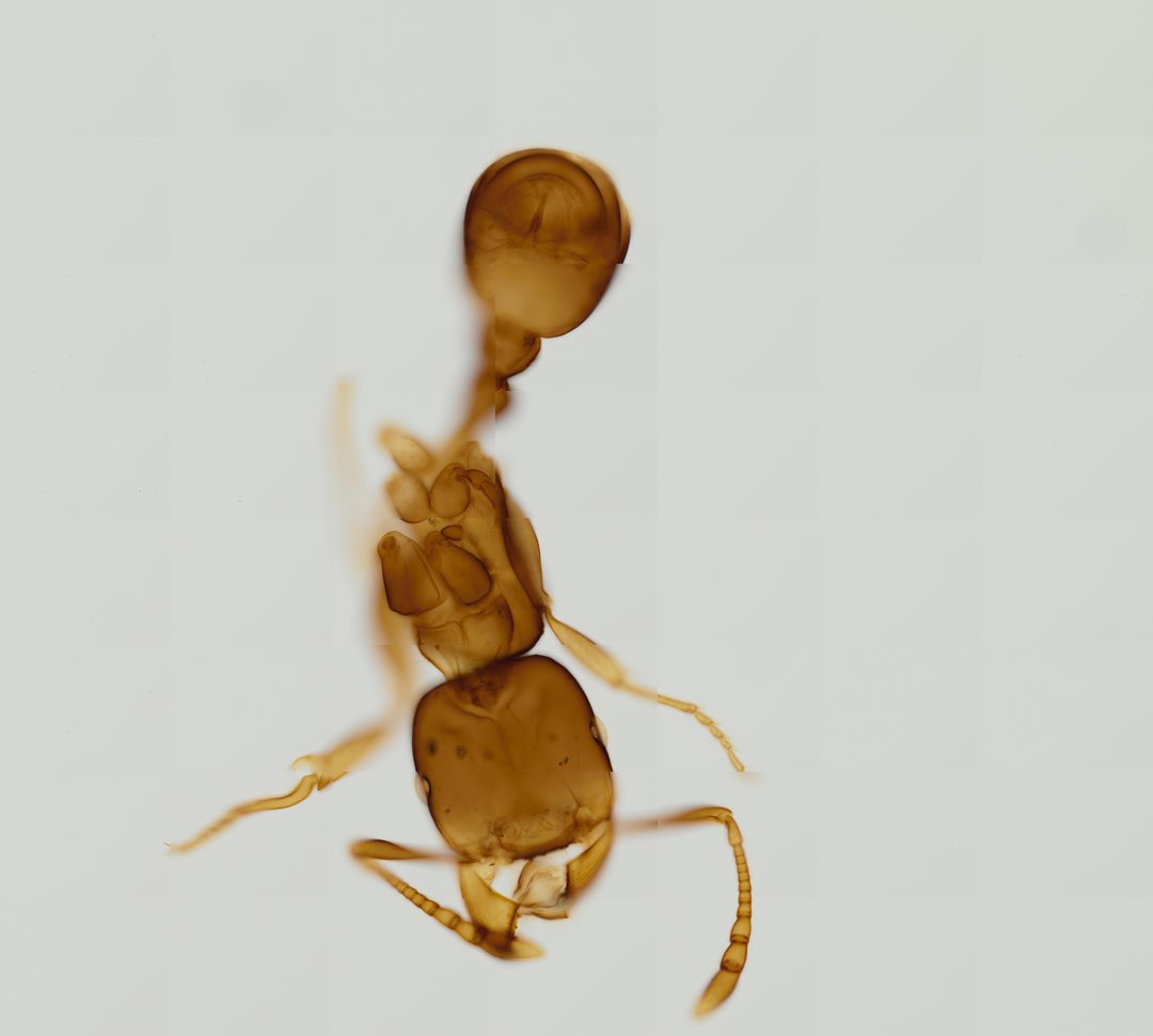
This is a whole ant mounted flat on a slide without stain, so you see its natural amber to brown exoskeleton. It is a good specimen for learning the basic body plan of insects and the features that make ants easy to recognize.
A whole mount (W.M.) preparation of an ant (labeled Ant W.M.). The specimen illustrates key hymenopteran anatomy including a large head with elbowed antennae, a distinct mesosoma (thorax), narrow petiole node, and rounded gaster (abdomen).
What to look for
- Head: A large front section carrying the eyes, jaws (mandibles), and antennae.
- Elbowed antennae: Antennae with a long first segment and a sharp bend, like an elbow, a classic ant feature.
- Mesosoma (thorax): The middle section where all six legs attach.
- Petiole: A narrow waist of one or two small nodes between the thorax and abdomen.
- Gaster (abdomen): The rounded rear section behind the petiole.
At each magnification
Low power (about 4x)
The whole ant, with its three main body regions and six legs.
Medium power (about 10x)
The bend in the antennae, the jointed leg segments, and the narrow petiole.
High power (about 40x)
Fine hairs on the exoskeleton and the teeth along the edges of the mandibles.
Questions and answers
What are the body parts of an ant under a microscope?
Like all insects, an ant has a head, a thorax (called the mesosoma in ants), and an abdomen, with six legs attached to the thorax. Ants also have elbowed antennae and a narrow waist called the petiole that sets them apart from most other insects.
Why does this ant slide look brown instead of stained?
Whole insect mounts are often left unstained because the exoskeleton already has natural color and shows its shape well. The amber to brown color comes from the hardened cuticle itself.
- Category
- Invertebrates
- Scan magnification
- 20x
- Stain
- Unstained
- Tags
- Ant W.M., Ant w.m., Ant whole mount, Ant, Formicidae, Hymenoptera
Viewing this slide in class
This is a scan of a real prepared slide, not a simulation or a drawing. On StudyScope you can pan from the whole slide down to individual structures, always in focus, and measure them in real micrometers with the calibrated ruler. Present it live and every student screen follows your view and annotations. Students join with a room code on Chromebooks, iPads, or any browser, with no accounts.