Daphnia Under the Microscope
Daphnia (Whole Mount), Daphnia
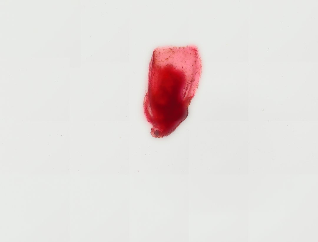
This is a whole Daphnia, or water flea, stained with carmine. Daphnia is a small see-through crustacean from ponds, and its clear body lets you see internal organs without cutting it.
This whole mount specimen of Daphnia, commonly known as a water flea, illustrates key morphological features of planktonic freshwater cladocerans. The body is enclosed in a transparent, bivalved chitinous carapace that leaves the head free. Prominent structures include a compound eye composed of fused ommatidia, a pair of large, branched second antennae utilized for jerky swimming locomotion, and a distinct digestive tract running centrally through the body cavity. Stained with carmine or eosin, internal organ structures including the brood chamber, heart, and thoracic filtration appendages (phylloids) are clearly delineated, offering insight into microscopic aquatic invertebrate physiology and filter-feeding mechanisms.
What to look for
- Carapace: A clear shell that wraps around the body but leaves the head free.
- Compound eye: A single dark eye near the front of the head.
- Second antennae: Large branched antennae used like oars for its jerky swimming.
- Digestive tract: A tube running through the body, often curved at the front.
- Heart: A small organ on the back, just behind the head.
- Brood chamber: A space on the back under the carapace where eggs or young may be held.
At each magnification
Low power (about 4x)
The whole oval body with the head, large antennae, and a spine at the rear.
Medium power (about 10x)
Internal organs showing through the carapace, stained pink to red by carmine.
High power (about 40x)
The compound eye, the bristles on the antennae, and the leaf-like legs inside the shell.
Questions and answers
What are the parts of a Daphnia under a microscope?
Daphnia has a clear carapace, a single compound eye, large branched antennae for swimming, a digestive tract, a heart on its back, and a brood chamber where eggs develop. Its legs are inside the carapace and are used for filter feeding.
Why is Daphnia used in biology labs?
Daphnia is transparent, so you can watch its heart beat and its gut work in living specimens. Classes often use live Daphnia to test how temperature or chemicals change heart rate.
- Category
- Invertebrates
- Scan magnification
- 20x
- Stain
- Carmine
- Tags
- Daphnia, water flea, Cladocera, whole mount, crustacean anatomy, carapace
Viewing this slide in class
This is a scan of a real prepared slide, not a simulation or a drawing. On StudyScope you can pan from the whole slide down to individual structures, always in focus, and measure them in real micrometers with the calibrated ruler. Present it live and every student screen follows your view and annotations. Students join with a room code on Chromebooks, iPads, or any browser, with no accounts.